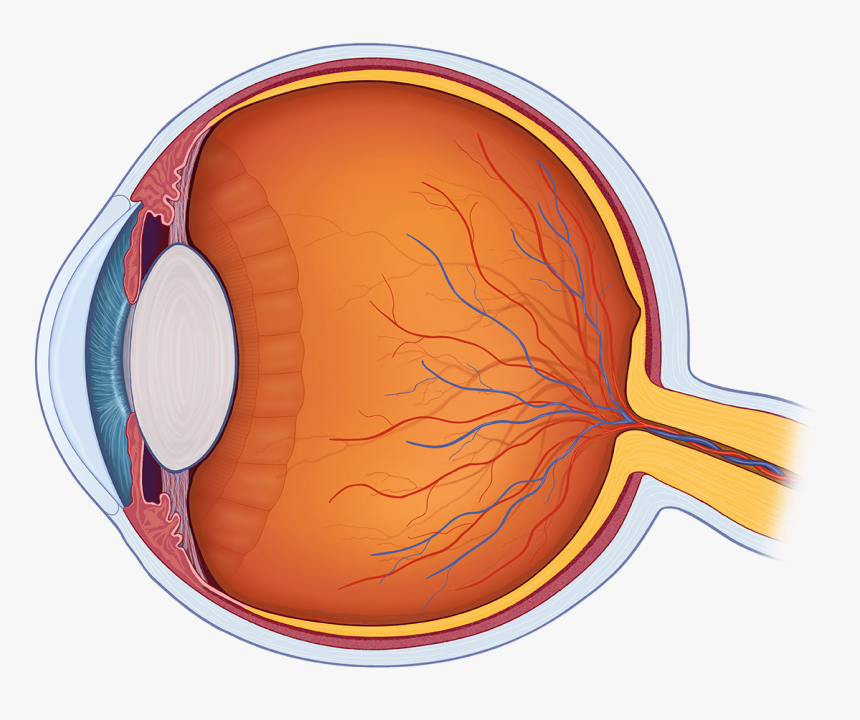
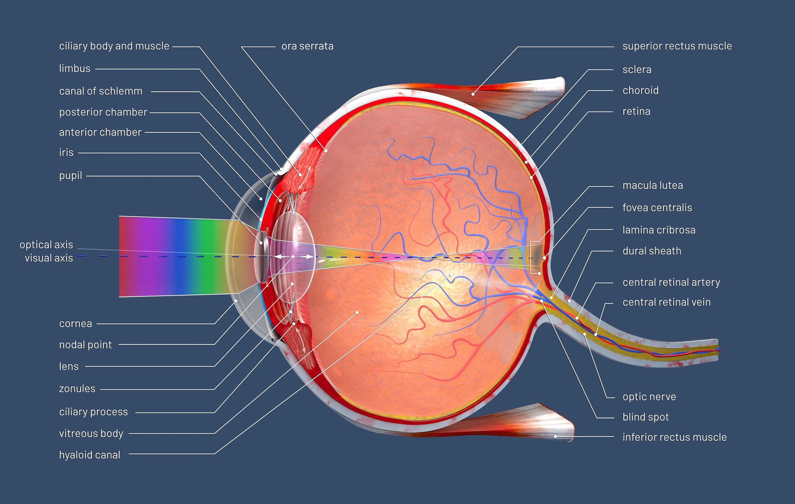
Eye in Cross Section. Click on a label to display the definition. Tap on the image or pinch out and pinch in to resize the image. On the cross-section of the eye, we can identify the two chambers of the eyeball filled with the aqueous humor; anterior and posterior. The anterior chamber of eyeball is found between the cornea and iris. The posterior chamber of eyeball is more of a slit-like cavity, found between the iris and lens. Fascial sheath (Tenon's capsule)

Human Eye Png Eye Ball Cross Section, Transparent Png , Transparent Png Image PNGitem
Cross-section of the eye. The zonules of Zinn keep the lens suspended, and the muscles of the ciliary body focus the lens. The ciliary body also secretes aqueous humor, which fills the anterior and posterior chambers, passes through the pupil into the anterior chamber, and drains primarily via the Schlemm canal. The iris regulates the amount of. Cross section of the human eyeball viewed from above. Dave Carlson / CarlsonStockArt.com. Iris. The choroid continues at the front of the eyeball to form the iris. The iris is a flat, thin, ring-shaped structure sticking into the anterior chamber. This is the part that identifies a person's eye colour. Cross-section of the eye The zonules of Zinn keep the lens suspended, and the muscles of the ciliary body focus the lens. The ciliary body also secretes aqueous humor, which fills the anterior and posterior chambers, passes through the pupil into the anterior chamber, and drains primarily via Schlemm's canal. Fig. 2. Sagittal section of the adult human eye. A cross-sectional view of the eye shows: Three different layers. The external layer, formed by the sclera and cornea; The intermediate layer, divided into two parts: anterior (iris and ciliary body) and posterior (choroid) The internal layer, or the sensory part of the eye, the retina

eye cross section Discovery Eye Foundation
A cross-sectional view of the eye shows : Three different layers: The external layer, formed by the sclera and cornea. The. The sagittal section of the eye also reveals the lens, which is a transparent body located behind the iris. The lens is suspended by ligaments (called zonule fibers) attached to the anterior portion of the ciliary body.. Cross-section of the eye. The zonules of Zinn keep the lens suspended, and the muscles of the ciliary body focus the lens. The ciliary body also secretes aqueous humor, which fills the anterior and posterior chambers, passes through the pupil into the anterior chamber, and drains primarily via Schlemm's canal. The iris regulates the amount of. Cross-section of the eye. The zonules of Zinn keep the lens suspended, and the muscles of the ciliary body focus the lens. The ciliary body also secretes aqueous humor, which fills the anterior and posterior chambers, passes through the pupil into the anterior chamber, and drains primarily via the Schlemm canal. The iris regulates the amount of. Representation of a horizontal section of the eyeball reveals its three coats: (1) external or fibrous coat (sclera and cornea); (2) middle or vascular coat (choroid, ciliary body, and iris); and (3) internal or retinal layer. The four refractive media are the cornea, the aqueous humor in the anterior chamber, the lens, and the vitreous body.

3d illustration of a cross section of the human eye with explanations and inscription Stephan
Ciliary body. The part of the eye that produces aqueous humor. Cornea. The clear, dome-shaped surface that covers the front of the eye. Iris. The colored part of the eye. The iris is partly responsible for regulating the amount of light permitted to enter the eye. Lens (also called crystalline lens). Dry eye syndrome is the most common eye disease, and if untreated, it can cause corneal ulcerations, scarring, and even perforation.. is an approximately 1.5mm area of specialized avascular retina that can be identified as a depression in the retina in cross-section. The foveola is the central floor of the fovea, approximately 0.35mm in.
Cross section of the eye. Note that the course of light travels through the cornea, pupil, and crystalline lens (becomes the cataract) to the retina and then to the brain through the optic nerve. An official website of the United States government Media in category "Human eyeball cross-section" The following 131 files are in this category, out of 131 total. 007alhazen eye.jpg 342 × 512; 38 KB. 1413 Structure of the Eye.jpg 2,175 × 1,242; 1.06 MB. 1490-95 da vinci - codex atlanticus.jpg 580 × 870; 157 KB. 201405 eye.png 400 × 400; 47 KB.

Human Eye Cross Section Eyeball 3D Model OBJ 3DS FBX C4D DXF STL
Behind the anterior chamber is the eye's iris (the colored part of the eye) and the dark hole in the middle called the pupil. Muscles in the iris dilate (widen) or constrict (narrow) the pupil to control the amount of light reaching the back of the eye. Directly behind the pupil sits the lens. The lens focuses light toward the back of the eye. Cross Section of an EyeballThe eyeball has three major coats-the smooth, protective outer sclera, the middle, pigmented choroid and the inner, light-sensitiv.


