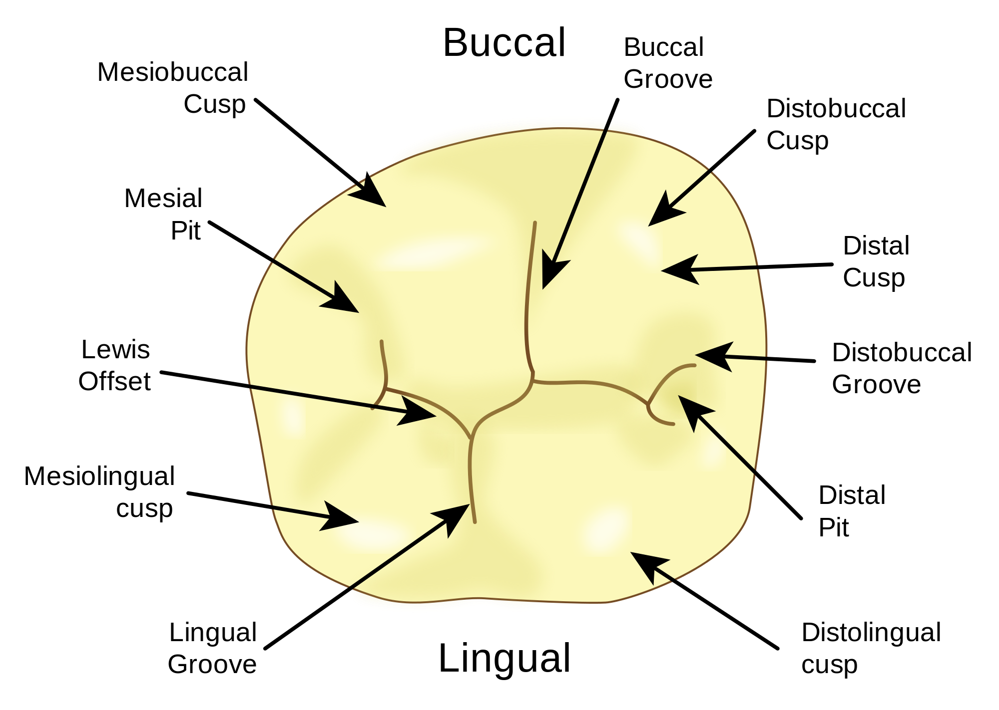
Labial - The surface towards the lips. Buccal - The surface towards the cheeks. Incisal - The biting edge of an anterior tooth. Lingual - The surface that faces the tongue. The surface of the tooth that is towards or adjacent to the tongue. The term lingual surface is used for the surface of a mandibular tooth (anterior or posterior) that is present immediately adjacent to the tongue. Clinical picture showing lingual and occlusal surfaces (Red arrows=Lingual surfaces) Mesial Surface

Your Tooth Surfaces Explained Dental Clinique
A chart is a diagrammatic representation of the teeth showing all the surfaces of the teeth. The charts in the examination will be used to show: Teeth present Teeth missing Work to be carried out Work completed Surfaces with cavities and restorations etc. When charting, the mouth is looked on as being a flat line. Labial Surface Diagram of the Tooth Numbering System (viewed as if looking into the mouth) Buccal (Facial) Surface Occlusal Surface Incisal Surface Right Left Maxillary Arch (Upper Jaw) Mandibular Arch (Lower Jaw) Adult Dentition = Permanent teeth 1-32 Child Dentition =Primary teeth A-T Wisdom Teeth =1, 16, 17, and 32 Dental charting is a process in which your dental healthcare professional lists and describes the health of your teeth and gums. Periodontal charting, which is a part of your dental. 1 Pronounce, define, and spell the Key Terms. 2 Describe what is included in a dental examination. 3 Describe Black's classification of cavities. 4 Identify charting symbols as related to dental needs and treatment. 5 Identify abbreviations used to name tooth surfaces. 6 Record a patient's existing condition.

Charting Surfaces Across Multiple Teeth at the Same Time Dentrix Enterprise Blog
What are the Surfaces of a Tooth? Learn the 5 Dental Surfaces Every Future Dentist Needs to KnowWhat are the surfaces of a tooth? Watch this video to learn t. Dental charting is a systematic method of recording vital information about a patient's oral health. Dentists use diagrams and symbols to document tooth identification, existing dental conditions, and various oral aspects or pathologies. This approach aids dentists in formulating tailored treatment plans and tracking patients' progress. Dental charting is the recording of a patient's dental structure and oral health to understand the condition of teeth and gums. It often involves using pictorial or graphic forms to provide a visual representation. The primary purpose of dental charting is to prepare detailed care plans for patients. With all the measurements on paper, you. The system involves numbering the labial/buccal surfaces as 1, the mesial surfaces as 2, the lingual surfaces as 3, the distal surfaces as 4, and the occlusal surfaces as 5. Anterior.

Here is a tooth chart (or a tooth map) that shows the lettering and numbering system that is
This measurement is made SIX times around each tooth for the standard charting. On the facial side of each tooth, it is measured close to the adjacent tooth on each side, and once in the middle.. This is also done on the lingual surface. For the measurements near the adjacent teeth it is a little tricky, because the tip of the probe must be. Week 4: Dental Charting. Welcome to week 4! This week you will spend some time reviewing earlier concepts about the teeth including surfaces and labelling, and learn about the dental chart. Dental charting combines information from week 2 (tooth labelling) and week 3 (dental conditions and procedures). Prior to moving forward, go back to weeks.
The tooth is one of the most individual and complex anatomical as well as histological structures in the body. The tissue composition of a tooth is only found within the oral cavity and is limited to the dental structures. Each tooth is paired within the same jaw, while the opposing jaw has teeth that are classified within the same category. However they are not grouped according to structure. Charting Terminology. Next>>. Apical - towards to the root. Buccal - surface of tooth towards cheeks. Coronal - towards the crown. Distal - surface away front midline. Facial - can be labial or buccal surface. Interproximal - surface between two teeth. Labial - surface of tooth towards lips.

Introduction to Dental Anatomy (Dental Anatomy, Physiology and Occlusion) Part 2
apical - Toward the root of the tooth; apex of the tooth. bifurcated - Single tooth with two roots. buccal - The surface that is facing the cheeks in the back of the mouth. cementum - The tissue covering the root of the tooth. cementoenamel junction (CEJ) - The line where the enamel and the cementum of the tooth join. Teeth numbers 1, 16, 17, and 32 are your wisdom teeth (3rd Molars). The teeth numbered 30 and 31 are your lower right molars. If you want to give your smile a new look with dental veneers (Fig. 4), your cosmetic dentist will enhance the most visible teeth in your mouth that always come out when you smile. These are teeth numbers 6 - 11 on the.


