Tailor your I.V. insertion techniques special populations; Manual and continuous bladder irrigation: Best practices; Managing your patient's arterial ulcer; Protecting your patient during a seizure; Related Links Articles in PubMed by SUE MASOORLI, RN; Decontaminate skin with alcohol 70% / chlorhexidine 2% swabs and leave to dry for at least 30 seconds. Use 'no-touch' technique for insertion after decontamination. Insert just distal to and along the line of the vein. Angle at 10-15° (Figure 2 below), or between 30-45° if using ultrasound guidance.
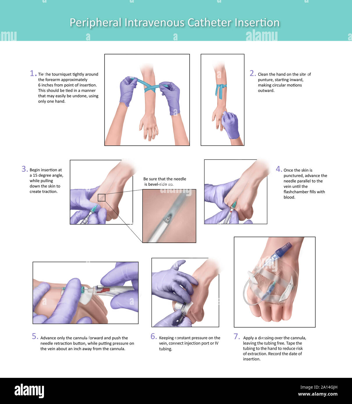
Illustration depicting the proper placement of a peripheral intravenous
Nurses who are deemed competent in IV insertion could continue to insert PIVC in consultation with NUM/CSN's. Definition of terms . A peripheral intravenous catheter (PIVC) is a thin plastic tube inserted into a vein using a needle. PIVCs allow for the administration of medications, fluids and/or blood products. Peripheral line placement, also referred to as peripheral intravenous (IV) cannulation, is the insertion of an indwelling single-lumen plastic conduit across the skin into a peripheral vein. Such devices may be referred to as peripheral IV (or venous) lines, cannulas, or catheters depending on the country. 2% alcoholic chlorhexidine, or 10% povidone iodine with 70% alcohol. Apply skin preparation solution meticulously to an area of skin approximately 10cm x 10cm, using concentric circles, for at least 30 seconds. Allow antiseptic to air dry completely. Do not palpate insertion site after antiseptic has been applied (unless it is re-prepped). Documentation Cues: Accurate and complete documentation regarding IV initiation should include the following: Description of the insertion site, such as "cephalic vein on dorsal surface of right lower arm, 2.5 cm (1 inch) above the wrist". Patient's tolerance of the procedure and patient education provided. If saline lock was established.

55 IV Therapy Tips and Tricks for Intravenous Nurses The Ultimate Guide
Sample Documentation of Unexpected Findings. Attempted to initiate IV infusion in right hand with existing 22-gauge IV catheter. IV site free from pain, redness, or signs of infiltration. IV site flushed readily with normal saline. Normal saline IV fluids connected at 200 mL/hour with immediate leaking around infusion site. Peripheral venous cannulation, among the most common medical procedures, has revolutionized the practice of medicine. Peripheral intravenous (IV) catheters allow for the safe infusion of medications, hydration fluids, blood products, and nutritional supplements. First-time success rate for peripheral IV placement ranges from 65 to 86 percent [ 1 ]. IV insertion equipment varies among institutions, but common types include shielded IV catheters or winged (i.e., "butterfly") devices. Variation is often related to the presence of a stabilizing device at the site of insertion, as well as the presence of short extension tubing.. This should be recorded in the patient's chart and is. Immediately remove the cannula, notify the provider, and document findings in the chart. In addition to local complications that can occur at the site of IV insertion, there are many systemic complications that nurses must monitor for when initiating peripheral IV access, as well as monitoring a client receiving IV therapy..

starting an iv line Mara Norwood
Get the IV line ready and set up the IV bag. Prepare your IV while your patient's arm (or other area of IV insertion) dries from the disinfectant wipe. Begin by preparing your IV tubing. Hang the IV bag from something elevated and fill the tubing with saline solution. Watch for any signs of bubbles in the IV line. IV Placement Chart showing veins of the arm and lower leg. Veins that are 'deeper' or those that you can't see usually provide better insertion sites than superficial veins which are mostly thin. However, try to avoid thick veins just below a bifurcation (i.e. a point where the thick vein branches out into smaller veins) as.
1. Disinfect the IV site. Next, tear open a fresh alcohol wipe (or use a similar sterilising method like chlorhexidine) and apply it to the skin in the area that the IV will be inserted. Wipe gently but thoroughly, ensuring an even coat of alcohol. Practice Criteria B - Documentation should include, but not be limited to, the following. 2. Type, brand, length, and size of vascular device. 3. Date and time of insertion, number and location of attempts, type of catheter stabilization and dressing, patient's response to the insertion, and identification of the person inserting the device.

How to Start an IV? 50+ Tips on IV Insertion, Rolling Veins (2020 Update)
Gravity. Putting the arm in a dependent position forces blood pooling in the distal veins, which will make them bigger and easier to see and palpate. This should make IV insertion easier with a higher chance of success. Also Read: "10 ER Nursing Hacks you Need to Know". The Management of Peripheral Intravenous Catheters Clinical Care Standard includes ten evidence-based quality statements to promote the skillful use of peripheral intravenous catheters (PIVCs) and to reduce complications including infections.. Insertion of a PIVC pre-emptively may be appropriate for patients at risk of clinical deterioration.
