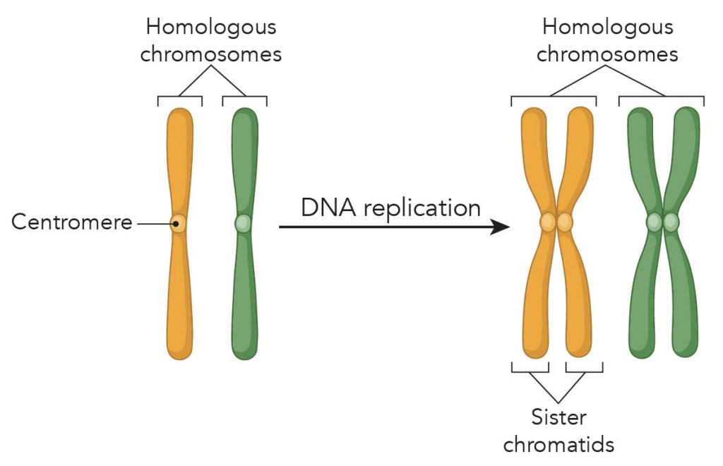
Chromosomes are a key part of the process that ensures DNA is accurately copied and distributed in the vast majority of cell divisions. Still, mistakes do occur on rare occasions. Changes in the number or structure of chromosomes in new cells may lead to serious problems. Introduction When a cell divides, one of its main jobs is to make sure that each of the two new cells gets a full, perfect copy of genetic material. Mistakes during copying, or unequal division of the genetic material between cells, can lead to cells that are unhealthy or dysfunctional (and may lead to diseases such as cancer).

Image result for structure of chromosome Chromosome, Chromosome
The process of differentiating between cells is expressed by labeling the developmental tree. Tracing a path from the root to a specific cell in the tree reveals the history of its divisions. Normal human fetal cells will divide approximately 40 to 60 times before cell division halts as demonstrated by Hayflick [ 1 ]. In this paper, we describe the labeling of human genomic loci in live cells with three orthogonal CRISPR/Cas9 components, allowing multicolor detection of genomic loci with high spatial resolution, which provides an avenue for barcoding elements of the human genome in the living state. Thoru Pederson Nature Biotechnology 34 , 528-530 ( 2016) Cite this article 35k Accesses 296 Citations 168 Altmetric Metrics Abstract A lack of techniques to image multiple genomic loci in living. < Prev Next > Chromosome Map Our genetic information is stored in 23 pairs of chromosomes that vary widely in size and shape. Chromosome 1 is the largest and is over three times bigger than chromosome 22. The 23rd pair of chromosomes are two special chromosomes, X and Y, that determine our sex.

What is Mitosis (Food model of mitosis) Rs' Science
A karyotype is the number and appearance of chromosomes. To obtain a view of an individual's karyotype, cytologists photograph the chromosomes and then cut and paste each chromosome into a chart, or karyogram, also known as an ideogram. In a given species, chromosomes can be identified by their number, size, centromere position, and banding. Figure 1 Construction of the CRISPR/Cas9 imaging system for fluorescent labeling of a particular chromosome in live cells. (A) Scatter plot for numbers of sgRNA-binding sites in each cluster of. chromosome, the microscopic threadlike part of the cell that carries hereditary information in the form of genes.A defining feature of any chromosome is its compactness. For instance, the 46 chromosomes found in human cells have a combined length of 200 nm (1 nm = 10 − 9 metre); if the chromosomes were to be unraveled, the genetic material they contain would measure roughly 2 metres (about 6. Chromosome number. Different species have different numbers of chromosomes. For example, humans are diploid (2n) and have 46 chromosomes in their normal body cells. These 46 chromosomes are organized into 23 pairs: 22 pairs of autosomes and 1 pair of sex chromosomes. The sex cells of a human are haploid (n), containing only one homologous.

Labeled Chromosome Structure Diagram imgprobe
In one application, by labeling and tracking the broken ends of chromosomal fragments, CRISPR FISHer enables real-time visualization of the entire process of chromosome breakage, separation, and. 1. INTRODUCTION Studies initiated during the last quarter of the nineteenth century gradually revealed the key roles of chromosomes in storing and transmitting hereditary information and in generating genetic variation. The most frequent method to study chromosome organization and behavior had been optical microscopy.
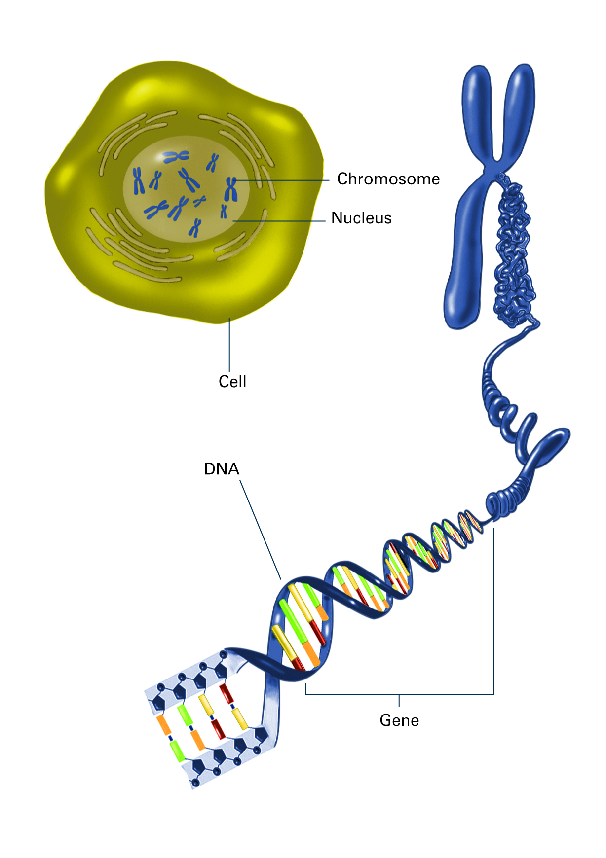
The first number or letter used to describe a gene's location represents the chromosome. Chromosomes 1 through 22 (the autosomes) are designated by their chromosome number. The sex chromosomes are designated by X or Y. The arm of the chromosome. Chromosome Definition. A chromosome is a string of DNA wrapped around associated proteins that give the connected nucleic acid bases a structure. During interphase of the cell cycle, the chromosome exists in a loose structure, so proteins can be translated from the DNA and the DNA can be replicated. During mitosis and meiosis, the chromosome.

Image and Video Gallery National Institute of General Medical Sciences
Chromosome Structure - A Simple Labeling Exercise Chromosome Structure This simple worksheet shows a diagram of a chromosome and where it is located in the nucleus of the cell. Students use a word bank to label the chromatid, centromere, chromosomes, cell membrane, DNA, and nucleus. Students label a simple diagram of a chromosome showing the centromere, chromatid, DNA, and the location of the chromosome within the nucleus of a cell.


