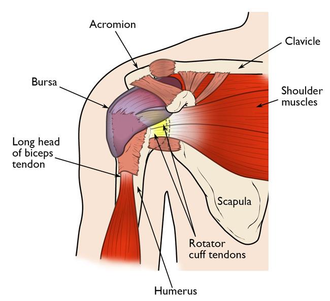
Description The rotator cuff is a common source of pain in the shoulder. Pain can be the result of: Tendinitis. The rotator cuff tendons can be irritated or damaged. Bursitis. The bursa can become inflamed and swell with more fluid causing pain. Impingement. Anatomy of the rotator cuff The rotator cuff comprises four tendons — the supraspinatus, infraspinatus, teres minor, and subscapularis; each of them attaches a muscle of the same name to the scapula (shoulder blade) and the humerus, or upper arm bone (see illustration).

Shoulder Joint Medial & Lateral Rotation In Abduction G4
A normal range of motion for shoulder extension to the highest point you can lift your arm behind your back — starting with your palms next to your body — is between 45 and 60 degrees. Shoulder. The Rotator Cuff (RC) is a common name for the group of 4 distinct muscles and their tendons, which provide strength and stability during motion to the shoulder complex. They are also referred to as the SITS muscle, with reference to the first letter of their names ( Supraspinatus, Infraspinatus, Teres minor, and Subscapularis, respectively). Acting in conjunction with the pectoral girdle, the shoulder joint allows for a wide range of motion at the upper limb; flexion, extension, abduction, adduction, external/lateral rotation, internal/medial rotation and circumduction. In fact, it is the most mobile joint of the human body. Action: Shoulder lateral rotation Nerves: Axillary and suprascapular Skeletal muscles: Deltoid, infraspinatus, and teres minor Cutaneous distribution: None except for the axillary nerve Neuromuscular deficit: Weakness/paralysis when rotating laterally at the shoulder joint under resistance.

Shoulder Impingement/Rotator Cuff Tendinitis OrthoInfo AAOS
The shoulder joint (glenohumeral joint) is an articulation between the scapula and the humerus. It is a ball and socket -type synovial joint, and one of the most mobile joints in the human body. In this article, we shall look at the anatomy of the shoulder joint - its structure, blood supply, and clinical correlations. Anatomical Structure In the human body, the rotator cuff is a functional anatomical unit located in the upper extremity . Its function is related to the glenohumeral joint, where the muscles of the cuff function both as the executors of the movements of the joint and the stabilization of the joint as well. Introduction. The rotator cuff is a group of muscles in the shoulder that allow a wide range of movement while maintaining the stability of the glenohumeral joint. The rotator cuff includes the following muscles [1] [2] [3] : A helpful mnemonic to remember these muscles is "SITS". The glenohumeral joint is a ball and socket joint and comprises. The shoulder is structurally and functionally complex as it is one of the most freely moveable areas in the human body due to the articulation at the glenohumeral joint. It contains the shoulder girdle, which connects the upper limb to the axial skeleton via the sternoclavicular joint. The high range of motion of the shoulder comes at the expense of decreased stability of the joint, and it is.

Humerus and Shoulder Joint
Other shoulder muscles. Pectoralis minor protects your shoulder blade and allows you to lower a shoulder. Latissimus dorsi is responsible for extension, adduction, and the medial rotation of your. Infraspinatus is the main muscle responsible for lateral rotation of your arm away from the centerline of your body. It's a thick triangular muscle. It covers the back of your shoulder blade.
36 Questions 3 Evidence 9 Video/Pods 28 Images Physical exam components This topic is broken down into Inspection Palpation Range of Motion Neurovascular Provocative tests Inspection Important to compare both shoulders skin scars symmetry swelling atrophy hypertrophy scapular winging Of all the movements that the shoulder can do, medial and external (also known as lateral) rotation are the most problematic. How shoulder rotation affects athletic performance . Shoulder strength and mobility can have a huge impact on athletic performance. Tight shoulder rotators will limit your range of motion.

SHOULDER EXTERNAL ROTATION LATERAL YouTube
Shoulder mobilization are a key examination tool to assess the integrity of accessory joint motion. Shoulder mobilizations, often utilized in manual therapy, also serve as a treatment procedure and are commonly administered in cases where joint range of motion is restricted. Specific grades of mobilization are described below. Medial and lateral rotation describe movement of the limbs around their long axis: Medial rotation is a rotational movement towards the midline. It is sometimes referred to as internal rotation. To understand this, we have two scenarios to imagine.


