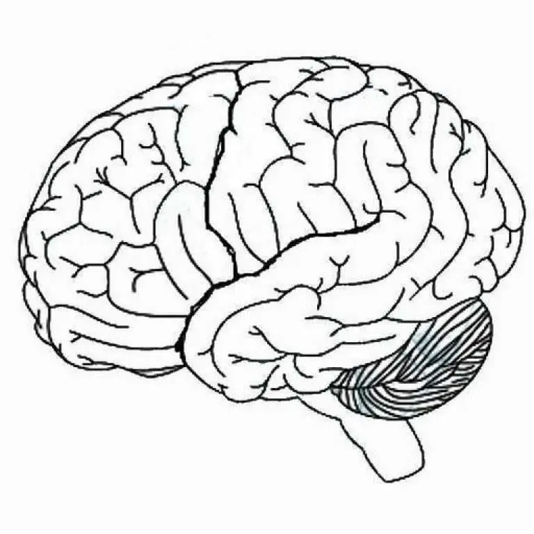
Next, here are 3 unlabeled human brain diagrams. Students can print these and practice labeling the parts of the human brain in preparation for a psychology, biology, or anatomy quiz or test. Blank Human Brain Diagram. Finally, here is a set of blank human brain diagrams. These may be useful for teachers to include in posters, presentations. Labeled brain diagram. First up, have a look at the labeled brain structures on the image below. Try to memorize the name and location of each structure, then proceed to test yourself with the blank brain diagram provided below.. Unlabeled (blank) diagram to be used as a worksheet Learn faster with quizzes. Learn all about brain structure.
Unlabeled Brain Diagram Cliparts.co
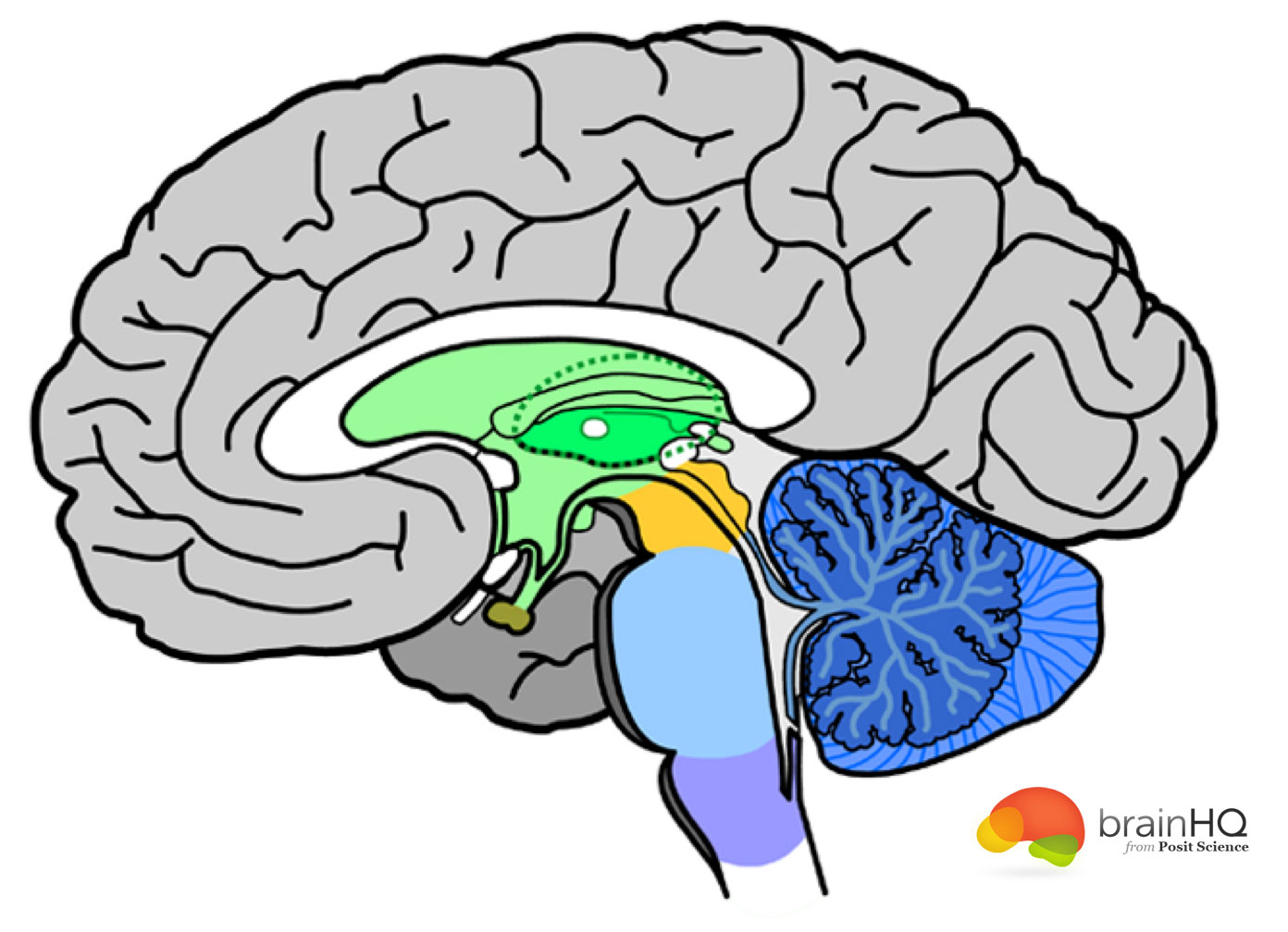
Illustrations and diagrams of the brain. These original illustrations and diagrams of the brain were created from 3D medical imaging reconstructions and then redrawn and colored using Adobe Illustrator. These anatomical charts include the main diagrams necessary for medical students, nursing students, residents, practitioners, anatomists to. Learning objective: to recognize the relationships between the embryological subdivisions of the brain and their derivatives that are visible in the adult brain.. Fifth Edition textbook). Once in Sylvius4 Online, go to Surface Anatomy, Photographic Atlas, and then click on either Unlabeled or Sulci and Fissures. Next, click on the thumbnail. The midsagittal section of the brain shows the three major parts of the brain, which are the cerebrum, cerebellum, and brainstem.The cerebrum (prosencephalon or forebrain) comprises the telencephalon (cerebral hemispheres) and the diencephalon.They are each also divided into subparts or regions for simplified localization of structures, for example, the brainstem is composed of the midbrain. What's In Your Brain? Activity Key 1. Cerebral cortex 2. Thalamus 3. Corpus callosum 4. Hypothalamus 5. Hippocampus 6. Pituitary gland 7. Midbrain 8. Pons 9. Medulla 10. Brainstem 11. Spinal cord 12. Cerebellum
Printable Blank Brain Diagram Printable World Holiday
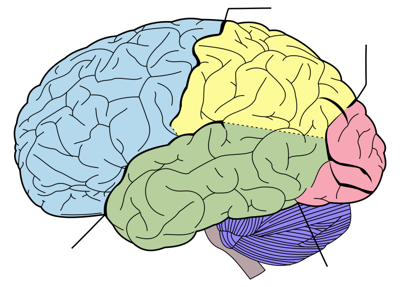
The lateral view of the brain shows the three major parts of the brain: cerebrum, cerebellum and brainstem.. A lateral view of the cerebrum is the best perspective to appreciate the lobes of the hemispheres. Each hemisphere is conventionally divided into six lobes, but only four of them are visible from this lateral perspective.The lobes are named after the bones of the skull that overlie them: Meninges of the brain, unlabeled diagram. Medical illustration of the layers of the meninges of the brain, very detailed. Brain stroke diagrams.. Diagram of visual projection pathway from eyes to brain (visual cortex), unlabeled. Referred pain chart, labeled diagram. Areas on body where pain from internal organs are perceived. Labeled drawing. In Part 2 of Brain Matters: Brain Diagram, students fill in the names of structures and functions on an unlabeled brain map. They check accuracy by comparing their work with other students. Parts 1 and 2 can be completed independently of each other. Goals and Objectives Students will be able to: • identify the functions of structures of the brain Image size: 31.2 Mpixels (89.4 MB uncompressed) - 6250x5000 pixels (20.8x16.6 in / 52.9x42.3 cm at 300 ppi)
Brain Diagram Unlabeled ClipArt Best
Human brain anatomy, unlabeled diagram. Median section of brain. Meninges of the brain. Meninges of the brain. Middle cerebral artery, labeled drawing. Middle cerebral artery and branches. Motor and sensory areas of the brain. Speech centers of the brain. Cortical language control updated. Brain Stem: two main parts - pons and medulla • Brain's most primitive part • Controls simple reflexes, such as coughing, sneezing, and digestion • Pons contains the fibers that connect the cerebral cortex with the cerebellum and spinal cord, and also controls sleep, awakening, and dream onset •
The anterior circulation. The four major arteries that arise from the internal carotid artery plus the posterior cerebral artery form the anterior circulation. The pattern of branching of each artery is similar: each gives rise to branches that supply cortical structures and each gives rise to branches that penetrate the ventral surface of the brain and supply deep structures (the basal. Try our top 10 quizzes : 1 - the skeleton: test your knowledge of the bones of the full skeleton. 2 - the brain: can you name the main anatomical areas of the brain?. 3 - the cell: learn the anatomy of a typical human cell. 4 - the skull: Do you know the bones of the skull?. 5 - the axial skeleton: How about the bones of the axial skeleton?. 6 - the heart: name the parts of the human heart
Printable Blank Brain Diagram Printable Word Searches
Our collection includes a variety of brain diagrams, ranging from simple and labeled diagrams for beginners, to more detailed and unlabeled diagrams for advanced learners. Each diagram highlights key structures and areas of the brain, such as the frontal lobe, parietal lobe, occipital lobe, temporal lobe, and cerebellum, among others. We introduce the Mindboggle-101 dataset, the largest and most complete set of free, publicly accessible, manually labeled human brain images. To manually label the macroscopic anatomy in magnetic resonance images of 101 healthy participants, we created a new cortical labeling protocol that relies on robust anatomical landmarks and minimal manual edits after initialization with automated labels.
