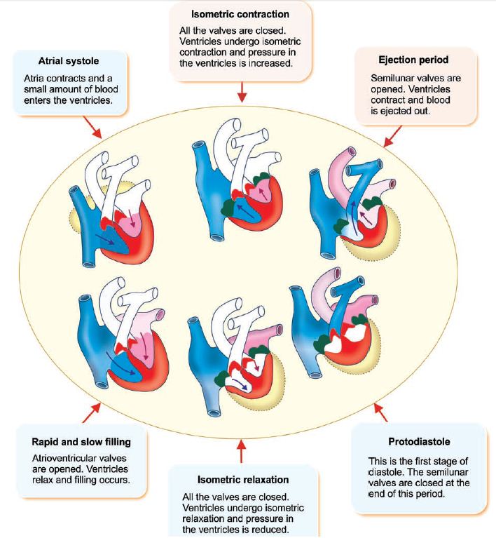
CARDIAC CYCLE The cardiac cycle is a period from the beginning of one heart beat to the beginning of the next one. It consists of two parts: Ventricular contraction called Ventricular relaxation called systole. diastole. Each part of the cardiac cycle consists of several phases characterized by either Cardiac Cycle Notes: Diagrams & Illustrations | Osmosis Exit full screen Full screen Cardiac Cycle Notes Contents Measuring cardiac output (Fick principle) Cardiac and vascular function curves Stroke volume, ejection fraction, and cardiac output Altering cardiac and vascular function curves Pressure-volume loops Changes in pressure-volume loops

Cardiac Cycle Physiology & Major Events PDF Download
Constructing the Wiggers diagram using core concepts: a classroom activity. The cardiac cycle is a series of pressure changes that take place within the heart. These pressure changes result in the movement of blood through different chambers of the heart and the body as a whole. Figure 19.3.1 - Overview of the Cardiac Cycle: The cardiac cycle begins with atrial systole and progresses to ventricular systole, atrial diastole, and ventricular diastole, when the cycle begins again. Correlations to the ECG are highlighted. Pressures and Flow Cardiovascular Anatomy and Physiology Notes Contents Cardiovascular system anatomy and physiology Normal heart sounds Abnormal heart sounds Lymphatic system anatomy and physiology Osmosis High-Yield Notes This Osmosis High-Yield Note provides an overview of Cardiovascular Anatomy and Physiology essentials. The cardiac cycle is defined as a sequence of alternating contraction and relaxation of the atria and ventricles in order to pump blood throughout the body. It starts at the beginning of one heartbeat and ends at the beginning of another. The process begins as early as the 4th gestational week when the heart first begins contracting.

Cardiac Cycle Nursing school survival, Cardiac cycle, Medical school
The cardiac cycle shall a series away pressure changes that take place at the heart. These pressure changes result in the movement off blood through different chambers of the heart and the g as a whole. Diese pressure changes originate as conductive electrochemical changes within an myocardium ensure result is the conical contraction of cardiac muscles. CARDIAC CYCLE The cardiac cycle consists of precisely timed electrical and mechanical events that are re - sponsible for rhythmic atrial and ventricu-lar contractions. Figure 2.1 displays the pres - sure relationships between the left-sided cardiac chambers during the normal cardiac cycle and serves as a platform for describing key events. Explain the events of the cardiac cycle. Define cardiac output and stroke volume. Distinguish among the types of blood vessels, their structures, and their functions. Identify the major arteries and veins of the pulmonary circuit as well as the areas they serve. Describe the hepatic portal system. KEY TERMS cps.med.ubc.ca

Bernal Studio The Art of Daniel Bernal Phases of the Cardiac Cycle
Cardiovascular and lymphatic systems make up the circulatory system a vast network of organs and vessels responsible for the flow of: The Lymphatic system Lymph Lymph nodes Lymph vessels Blood Nutrients Hormones Oxygen and other gases To and from the Cells of the body The Cardiovascular system Blood Blood vessels Heart Cardiac Cycle. The cardiac cycle comprises a complete relaxation and contraction of both the atria and ventricles, and lasts approximately 0.8 seconds. Beginning with all chambers in diastole (relaxation), blood flows passively from the veins into the atria and past the atrioventricular valves into the ventricles. The atria begin to contract.
Human Anatomy & Physiology: Cardiovascular Physiology Ziser 2404 Lecture Notes, 2005 3 idea of how rapidly the impulses are being conducted and how the heart is functioning Cardiac Cycle 1 complete heartbeat (takes ~ 0.8 seconds) consists of: systole contraction of each chamber diastole relaxation of each chamber two atria contract simultaneously in a specialised area of cardiac cells, the sinoatrial node (SAN), situated in the right atrium. This is the heart's natural pace-maker. When working properly, it sets the heart rhythm (sinus rhythm) and initiates impulses that act on the myocardium, stim - ulating cardiac contraction. The cardiac impulse passes from the SAN into the atria,

Anatomy and Physiology Cardiac Cycle Notes Steps of the Etsy
The Cardiac Cycle consists of Diastole and Systole During diastole → heart relaxes and fills with blood During systole → the heart contracts and eject blood (i.e. emptying) Note: If heart rate is 72 beats/min, the duration of the cardiac cycle is about 0.8 second per beat. Of which 0.3 second is for systole and 0.5 second is for diastole. Wigger's diagram is used to demonstrate the varying pressures in the atrium, ventricle, and artery during one cardiac cycle (Figure 2). Intracardiac pressures are different within the right and left sides of the heart. The left side has higher pressure, as it has to pump blood through the whole body, compared to the right side, which has to.


