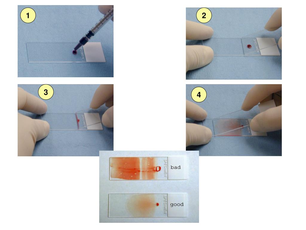
2. INTRODUCTION • Peripheral blood smear is a very important tool in the hematology lab • It provides rapid, reliable access to information about a variety of hematologic disorders • Examination of the peripheral blood smear is an inexpensive but powerful diagnostic tool in both children and adults • The smear offers a window into the functional status of the bone marrow • Review of. Place a drop of blood, about 2-3 mm in diameter approximately 1 cm from one end of slide. 2. Place the slide on a flat surface, and hold the other end between your left thumb and forefinger. 3. With your right hand, place the smooth clean edge of a second (spreader) slide on the specimen slide, just in front of the blood drop.

Peripheral Blood Smear Staining Procedure
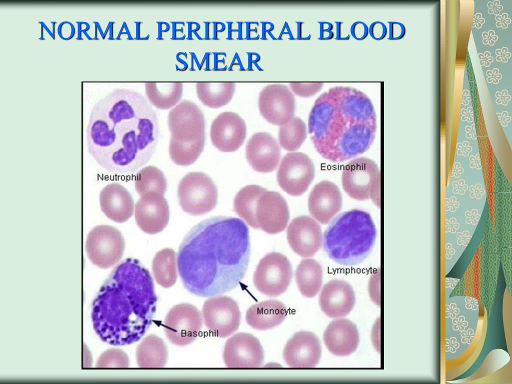
Examination of the peripheral blood smear is an inexpensive but powerful diagnostic tool in both children and adults. In some ways, it is becoming a "lost art," but it often provides rapid, reliable access to information about a variety of hematologic disorders. The smear offers a window into the functional status of the bone marrow, the. The diagnostic importance of a peripheral blood smear (PBS) is invaluable. A simple easy to do investigation can give invaluable information's of the various diseases. The PBF exposes the morphology of peripheral blood cells, which maintains its place in the morphologic diagnosis of various blood related diseases. Peripheral smear. 1. PERIPHERAL SMEAR. 2. PERIPHERAL SMEAR The role Examination of a well-made peripheral blood smear is a) Estimate approximately the numbers of each of the three cellular elements Red blood cells White blood cells Platelets b)Study the morphology of these c)See blood parasites. 3. Peripheral Smear Preparation • 22 x 27mm clean coverslip • More routinely used for bone marrow aspirate • Technique: 1. A drop of marrow aspirate is placed on top of 1 coverslip 2. Another coverslip is placed over the other allowing the aspirate to spread. 3. One is pulled over the other to create 1 thin smears.

PPT Peripheral Smear PowerPoint Presentation, free download ID4385589
Save figures into PowerPoint; Download tables as PDFs; Go to My Dashboard Close.. It is useful to ask the laboratory to generate a Wright's-stained peripheral blood smear and examine it. + + The best place to examine blood cell morphology is the feathered edge of the blood smear where red cells lie in a single layer, side by side, just. A note from Cleveland Clinic. A peripheral blood smear test is a technique healthcare providers use to examine your red and white blood cells and your platelets. This test gives them a clear picture of changes in your blood cells and platelets that may be a sign of disease. A peripheral blood smear test is an important part of diagnosing disease. Make a thin film of blood in the shape of tongue. Precautions: • Too slow a slide push will accentuate poor leukocyte distribution, larger cells are pushed at the end of the slide • Maintain an even gentle pressure on the slide • Keep the same angle all the way to the end of the smear. 16. The examination of a peripheral blood smear is one of the most informative exercises a physician can perform. Although advances in automated technology have made the examination of a peripheral blood smear by a physician seem less important, the technology is not a completely satisfactory replacement for a blood smear interpretation by a trained medical professional who also knows the patient.

PPT Peripheral blood smear examination PowerPoint Presentation, free download ID4893119
Peripheral Smear Preparation. Precautions: Ensure that the whole drop of blood is picked up and spread. Too slow a slide push will accentuate poor leukocyte distribution, larger cells are pushed at the end of the slide. Maintain an even gentle pressure on the slide. Keep the same angle all the way to the end of the smear. Peripheral Smear Preparation • Procedures: • Drop 2-3 mm blood at one end of the slide Diff safe can be used a. Easy dropping b. Uniform drop. The pusher slide be held securely with the dominant hand in a 30-45 deg angle. - quick, swift and smooth gliding motion to the other side of the slide creating a wedge smear.
Enough stain to cover entire smear-Enough stain to cover entire smear- keep for 2 min - fixation bykeep for 2 min - fixation by methanolmethanol 2.2. Add distilled water - double theAdd distilled water - double the amount—10 min -actual stainingamount—10 min -actual staining 3.3. Keep for 7 - 10 minKeep for 7 - 10 min 4.4. A well made peripheral smear is thick at one end and progressively thinner at the opposite end. The "zone of morphology" (area of optimal thickness for light microscopic examination) should be at least 2 cm in length. The smear should occupy the central area of the slide and be margin-free at the edges. PBS examination requires a systematic.

PPT Normal Red Blood Cells Peripheral Blood Smear PowerPoint Presentation ID345635
Peripheral Smear. An Image/Link below is provided (as is) to download presentation Download Policy: Content on the Website is provided to you AS IS for your information and personal use and may not be sold / licensed / shared on other websites without getting consent from its author. Download presentation by click this link. They are all artistically enhanced with visually stunning color, shadow and lighting effects. Many of them are also animated. And they're ready for you to use in your PowerPoint presentations the moment you need them. - PowerPoint PPT presentation. Peripheral Blood Smear Morphology William F. Kern, MD Director, Laboratory Hematology.


