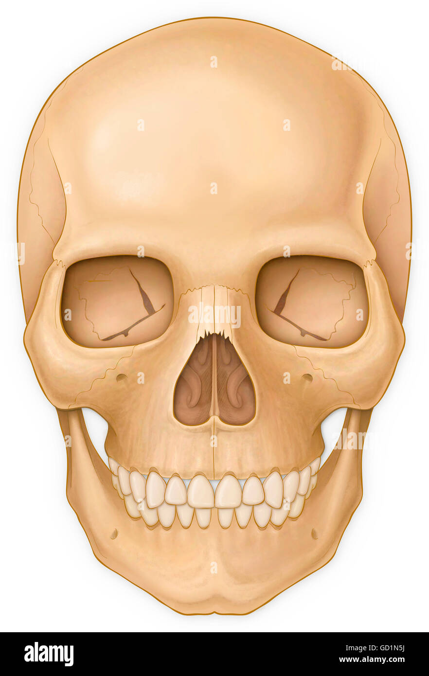
1/2 Synonyms: none The human skull consists of 22 bones (or 29, including the inner ear bones and hyoid bone) which are mostly connected together by ossified joints, so called sutures. The skull is divided into the braincase ( neurocr anium) and the facial skeleton ( viscerocranium ). The skull is the skeletal structure of the head that supports the face and protects the brain. It is subdivided into the facial bones and the cranium, or cranial vault ( Figure 7.3.1 ). The facial bones underlie the facial structures, form the nasal cavity, enclose the eyeballs, and support the teeth of the upper and lower jaws.

The Frontal View of the Skull Stock Illustration Illustration of skeleton, medically 162289174
Human Skull Front View Photos and Premium High Res Pictures - Getty Images Browse Getty Images' premium collection of high-quality, authentic Human Skull Front View stock photos, royalty-free images, and pictures. Human Skull Front View stock photos are available in a variety of sizes and formats to fit your needs. Browse Boards AI Generator 1/7 Synonyms: none The human skull consists of about 22 to 30 single bones which are mostly connected together by ossified joints, so called sutures. The skull is divided into the braincase ( cerebral cranium) and the face ( visceral cranium ). The main task of the skull is the protection of the most important organ in the human body: the brain. Human skull front view. Human skull 3d illustration. Front view on black background. Human skull in different angles. Isolated on black background. Side and front views. Anatomy and medicine concept. Skull Computer artwork of a wireframe view of a human skull Browse 4,091 skull front view photos and images available, or search for dog skull front view to find more great photos and pictures. Browse Getty Images' premium collection of high-quality, authentic Skull Front View stock photos, royalty-free images, and pictures.
Normal front view of the adult skull Stock Photo Alamy
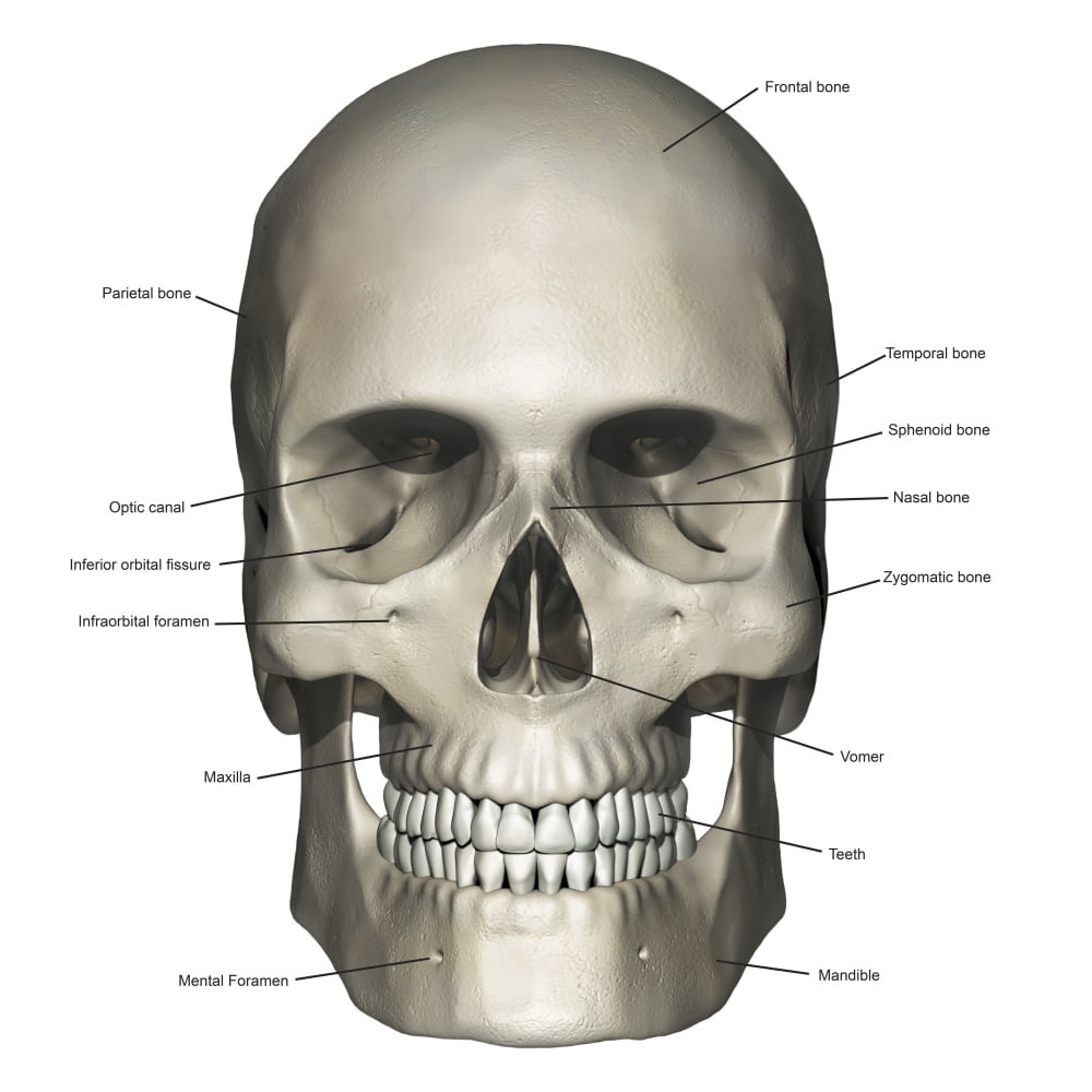
Figure 1. Parts of the Skull. The skull consists of the rounded brain case that houses the brain and the facial bones that form the upper and lower jaws, nose, orbits, and other facial structures. Watch this video to view a rotating and exploded skull, with color-coded bones. Frontal bone: the bone that. Temporal bone: the bones that form the inside of the sides of the skull and contain the zygomatic processes (the cheekbone), external auditory meatus. Crystal skull on dark background - Stock image. Crystal skull on dark background - Front view. Eagle and human skull. Black and white tattoo. Eagle sitting on the human skull. Black and white Tattoo or shirts design style vector illustration. Front view. Set of the vector skulls isolated on white background. Browse Getty Images' premium collection of high-quality, authentic Skull Front View stock photos, royalty-free images, and pictures. Skull Front View stock photos are available in a variety of sizes and formats to fit your needs.
Anterior view of human skull anatomy with annotations Poster Print by Photon
Find Front View Skull stock images in HD and millions of other royalty-free stock photos, illustrations and vectors in the Shutterstock collection. Thousands of new, high-quality pictures added every day. skull, skeletal framework of the head of vertebrates, composed of bones or cartilage, which form a unit that protects the brain and some sense organs. The upper jaw, but not the lower, is part of the skull. The human cranium, the part that contains the brain, is globular and relatively large in comparison with the face.
The human skeletal system, vector illustrations of human skeleton front and rear view. Eagle and human skull. Black and white tattoo. Eagle sitting on the human skull. Black and white Tattoo or shirts design style vector illustration. Front view. The front view of skull almost brings into view all the bones that make up the skull, with a few exceptions. The different bones viewed from the front of sku.
Human Skull 2 Free Stock Photo Public Domain Pictures
Head anatomy Author: Adrian Rad BSc (Hons) • Reviewer: Dimitrios Mytilinaios MD, PhD Last reviewed: October 30, 2023 Reading time: 7 minutes Recommended video: Muscles of mastication [22:28] Origins, insertions, innervation and functions of the muscles of mastication. Human head (anterior view) Technical factors posteroanterior projection centering point the beam is exiting at the nasion collimation superior to the skin margins inferior to include the most inferior aspects of the skull lateral to include the skin margin orientation portrait detector size 24 cm x 30 cm exposure 75-80 kVp 20-25 mAs SID 100 cm grid
