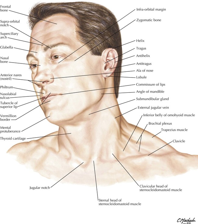
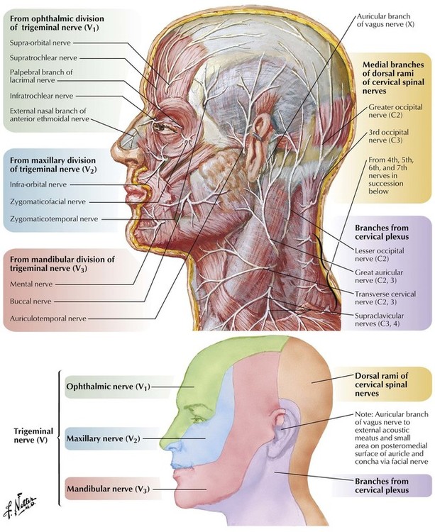
muscular triangle contains the strap muscles of the neck; larynx and thyroid gland lie deep to the strap muscles: omoclavicular triangle: boundaries: superior - inferior belly of the omohyoid m.; anterior - sternocleidomastoid m.; inferior - middle 1/3 of the clavicle: the external jugular vein courses deeply through the omoclavicular triangle Larynx. Salivary glands. Motor and sensory innervation of face and oral cavity. Lymphoid system of face and neck. Coniotomy, tracheotomy. Term ANATOMY from Greek anatomé = „a cutting up" SYSTEMATIC ANATOMY As a science deals with morphology and structures of the human body.

1 Head and Neck Basicmedical Key
Skull Nose and nasal cavity Eye Ear Mouth Tooth Neck Sources Related articles + Show all Skull The skull is a strong, bony capsule that rests on the neck and encloses the brain. It consists of two major parts: the neurocranium (cranial vault) and the viscerocranium (facial skeleton). Head Caput 1/2 Synonyms: none The head is the superior part of the body that is attached to the trunk by the neck. It is the control and communication center as well as the "loading dock" for the body. It houses the brain and therefore is the site of our consciousness: ideas, creativity, imagination, responses, decision making and memory. The deep cervical fasciae of the neck, since its first description in the early 1800s, have been a source of considerable controversy amongst anatomists. Several different classification systems for these anatomic structures have been proposed based on topographic morphology, embryologic origin, and surgical approach. The most commonly accepted classification system found in the literature is. Written by an experienced and well-respected physician and professor, this new volume, building on the previous volume, Ultrasonic Topographical and Pathotopographical Anatomy, also available from Wiley-Scrivener, presents the ultrasonic topographical and pathotopographical anatomy of the head and neck, offering further detail into these important areas for use by medical professionals.

Introduction in topographic anatomy and operative surgery презентация онлайн
Introduction The neck is the bridge between the head and the rest of the body. It is located in between the mandible and the clavicle, connecting the head directly to the torso, and contains numerous vital structures. Topographical Anatomy of the Head and Neck. Veins of the Head and Neck. Viscera of the Head and Neck. UAMS College of Medicine University of Arkansas for Medical Sciences. Mailing Address: 4301 West Markham Street, Little Rock, AR 72205. Phone: (501) 686-7000. Facebook; Twitter; Written by an experienced and well-respected physician and professor, this new volume, building on the previous volume, Ultrasonic Topographical and Pathotopographical Anatomy, also available from Wiley-Scrivener, presents the ultrasonic topographical and pathotopographical anatomy of the head and neck, offering further detail into these important areas for use by medical professionals. This. The medical information on this site is provided as an information resource only, and is not to be used or relied on for any diagnostic or treatment purposes.

1 Head and Neck Basicmedical Key
The triangles of the neck are the topographic areas of the neck bounded by the neck muscles. The s ternocleidomastoid muscle divides the neck into the two major neck triangles; the anterior triangle and the posterior triangle of the neck, each of them containing a few subdivisions. Abstract This chapter contains sections titled: Topographic Anatomy of the Neck Fasciae, Superficial and Deep Cellular Spaces and their Relationship with Spaces Adjacent Regions (Fig. 37) Triangles.
The movement of the head is mostly owing to its articulation with the neck. It acts as a bridge between the body and the head. Due to their proximity and directly linked functions, the head and neck regions are mentioned together. The head and neck areas are crucial to swallowing function. Therefore, the head provides the realization of many. This is a new edition of the classical topographical human anatomy by Pernkopf which is now offered as a two-volume atlas. Volume I is concerned with the head and neck. This volume consists of superbly executed anatomical illustrations most of which are in color. All of them are examples of high-quality engraving.

Anatomy of the Neck TrialExhibits Inc.
The roentgenograms of the skull and of the vertebral column of the head and neck skeleton and the arteriograms of the blood vessels of the neck and brain are vividly informative. The libraries of the medical schools will undoubtedly possess a copy of this book, yet it may be urged that all students. Spector B. Atlas of Topographical and. Anatomists will remember the second edition, published in 1980, as a premier topographical atlas of head and neck anatomy. T h i s new edition has main- tained the oversize format with single plates per page, but has added 67 color plates (298 total) and 10 black and white illustrations (1 18 total). Most of the drawings are based on cadaver.


