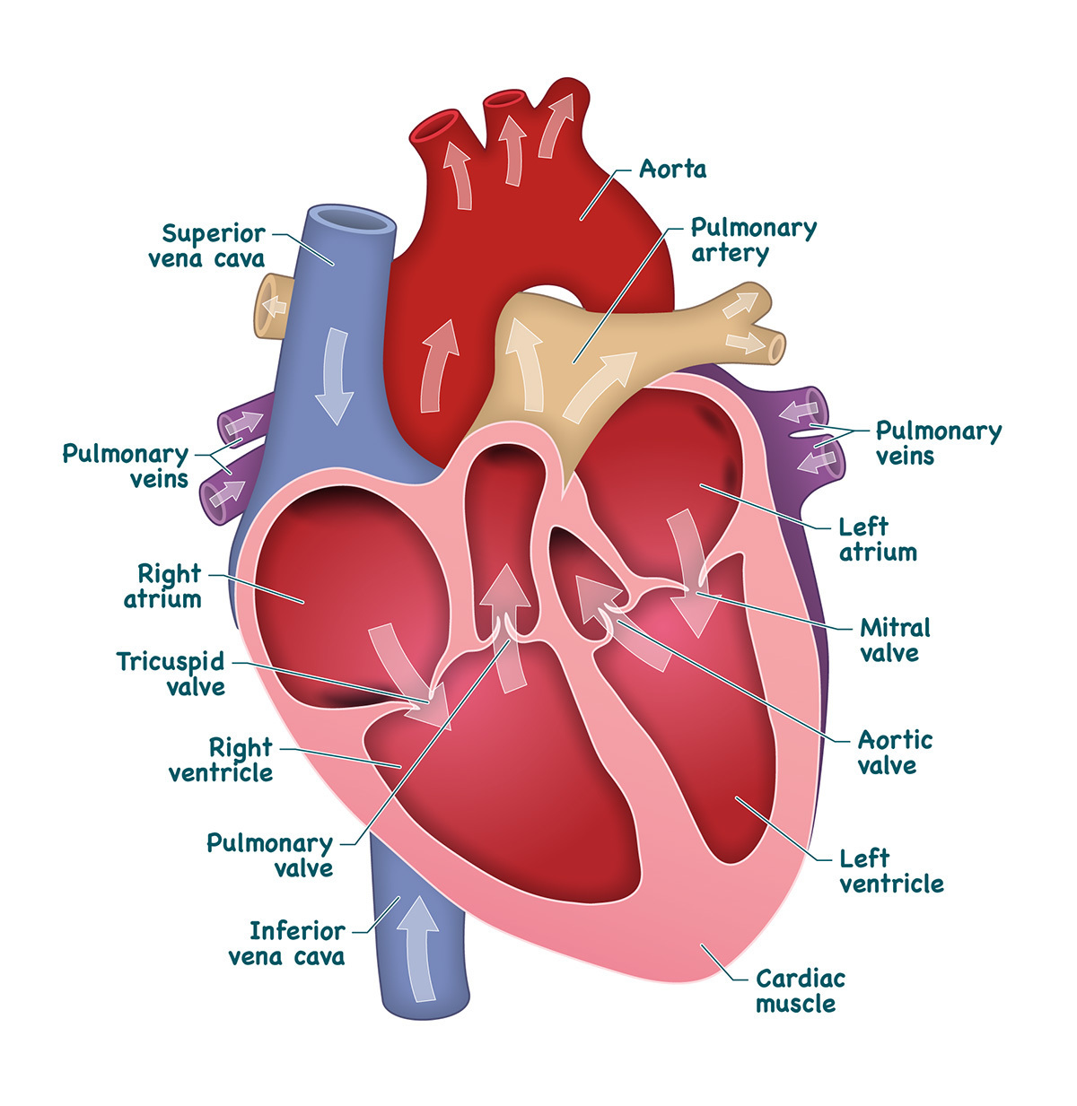
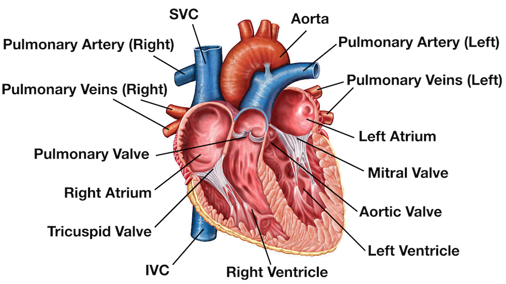
In this interactive, you can label parts of the human heart. Drag and drop the text labels onto the boxes next to the diagram. Selecting or hovering over a box will highlight each area in the diagram.. Drag and drop the text labels onto the boxes next to the heart diagram. If you want to redo an answer, click on the box and the answer will. The cusps are pushed open to allow blood flow in one direction, and then closed to seal the orifices and prevent the backflow of blood. Backward prolapse of the cusps is prevented by the chordae tendineae-also known as the heart strings-fibrous cords that connect the papillary muscles of the ventricular wall to the atrioventricular valves.. There are two sets of valves: atrioventricular.

Heart And Labels Drawing at GetDrawings Free download
The diagram of heart is beneficial for Class 10 and 12 and is frequently asked in the examinations. A detailed explanation of the heart along with a well-labelled diagram is given for reference. Well-Labelled Diagram of Heart. The heart is made up of four chambers: The upper two chambers of the heart are called auricles. Heart conditions are among the most common types of disorders affecting people. In the United States, heart disease is the leading cause of death for people of all genders and most ethnic and racial groups. Common conditions that affect your heart include: Atrial fibrillation (Afib): Irregular electrical impulses in your atrium. heart, organ that serves as a pump to circulate the blood.It may be a straight tube, as in spiders and annelid worms, or a somewhat more elaborate structure with one or more receiving chambers (atria) and a main pumping chamber (ventricle), as in mollusks. In fishes the heart is a folded tube, with three or four enlarged areas that correspond to the chambers in the mammalian heart. Function and anatomy of the heart made easy using labeled diagrams of cardiac structures and blood flow through the atria, ventricles, valves, aorta, pulmonary arteries veins, superior inferior vena cava, and chambers. Includes an exercise, review worksheet, quiz, and model drawing of an anterior vi

Show me a diagram of the human heart? Here are a bunch! Interactive Biology, with Leslie Samuel
The heart is made of three layers of tissue. Endocardium is the thin inner lining of the heart chambers and also forms the surface of the valves.; Myocardium is the thick middle layer of muscle that allows your heart chambers to contract and relax to pump blood to your body.; Pericardium is the sac that surrounds your heart. Made of thin layers of tissue, it holds the heart in place and. The user can show or hide the anatomical labels which provide a useful tool to create illustrations perfectly adapted for teaching. Anatomy of the heart: anatomical illustrations and structures, 3D model and photographs of dissection. Heart - Human anatomy : Cross sections (Right/left ventricle, Right/left atrium, Interventricular septum English: Heart diagram with labels in English. Blue components indicate de-oxygenated blood pathways and red components indicate oxygenated blood pathways. Date: March 2010: Source: Own work. Supporting references; The epicardium covers the heart, wraps around the roots of the great blood vessels, and adheres the heart wall to a protective sac. The middle layer is the myocardium. This strong muscle tissue powers the heart's pumping action. The innermost layer, the endocardium, lines the interior structures of the heart. 2.

Heart Anatomy Labeled Diagram, Structures, Blood Flow, Function of Cardiac System — EZmed
The heart is a muscular organ that pumps blood through the blood vessels of the circulatory system. Blood transports oxygen and nutrients to the body.. In this activity, students use online and paper resources to identify and label the main parts of the heart. By the end of this activity, students should be able to: The heart is divided into four chambers: two atria and two ventricles. Blood is transported through the body via a complex network of veins and arteries. The average human heart weighs between 6.
English: Heart diagram with labels in English. Blue components indicate de-oxygenated blood pathways and red components indicate oxygenated blood pathways. Date: March 2010: Source: Own work. Supporting references Blood flow through the heart made easy with a simple diagram of the cardiac circulation pathway and steps in order. Heart anatomy, video, quiz, and chart included! Great for USMLE, nursing, students, doctors, and medical learners.. you will be able to label the entire diagram shown below! The video above also provides an animation at the end.

OpenStax Anatomy and Physiology CH19 THE CARDIOVASCULAR SYSTEM THE HEART Top Hat Heart
If you are confused about where parts of the heart are, find a new diagram. Advertisement. Part 2. Part 2 of 3: Sketching the Heart. After you've drawn the structure, color the different sections of the heart distinct colors and appropriately label them. Make sure to write "The Human Heart" above your drawing as a title when you're. Higher; Structure and function of the heart The structure of the heart. In this Higher Human Biology revision guide, you will learn in detail that cardiac output is a measure of the rate of blood.


