Awesome prices & high quality here on Temu. New users enjoy free shipping & free return. Come and check all categories at a surprisingly low price, you'd never want to miss it. A viral image depicts a cat's tongue when imaged under a scanning electron micrograph. Rating: True About this rating Ever wondered what a cat's tongue looks like under a microscope?.
Why Does Your Cat's Tongue Feel Like Sandpaper? Deep Look KQED Science
A tongue under a microscope shows the below-mentioned histological features - Nonkeratinized stratified squamous epithelium on the tongue mucous membrane, Different types of papillae - filiform, fungiform, circumvallate, and foliate, Lamina propria layer in the mucosa membrane of the tongue, Here's What A Cat's Tongue Looks Like Under A Microscope Beyond the Press, a run by Finnish couple Lauri and Anni Vuohensilta, has wowed their over 700,000 subscribers with up-close, macro footage of their cats' tongues. The footage shows off the strange texture and hidden beauty of a difficult-to-capture part of the feline family. A composite image of a section of cat tongue plated on a microscope slide, dating back to the 1890s. The yellow streaks are horizontal muscles, the sparse purple streaks are vertical muscles, and the black squiggles are capillaries. Those blue-purple spots on the tongue's serrated edge represent polarized light interacting with keratin. On our channel we will show you everything that surrounds us under a microscope.An approximate list of what we will consider:- food under a microscope- water.
Cat Tongue Papillae Photograph by Dennis Kunkel Microscopy/science Photo Library Fine Art America
Once they had the tongues, each was scanned using micro-CT, which gives a detailed view of microscopic structures. The vestibule of a cat's oral cavity is a space between the internal surface of the lip and external to the teeth. Again, the mouth cavity proper of a cat extends from the inner surface of the teeth to the oropharynx. So, in the cat mouth cavity proper, you will find the gums, teeth, tongue, palates (hard and soft), and salivary glands. Science Mar 1, 2017 11:00 AM EST Even after thousands of years sharing our homes, cats still remain mysterious. For one thing, they spend an inordinate amount of time grooming themselves, up to. Magnify the cat's tongue 100 million times, Intricacies Unveiled: The Fascinating Realm of Microscopic Discovery

Cat tongue surface, SEM Stock Image Z934/0726 Science Photo Library
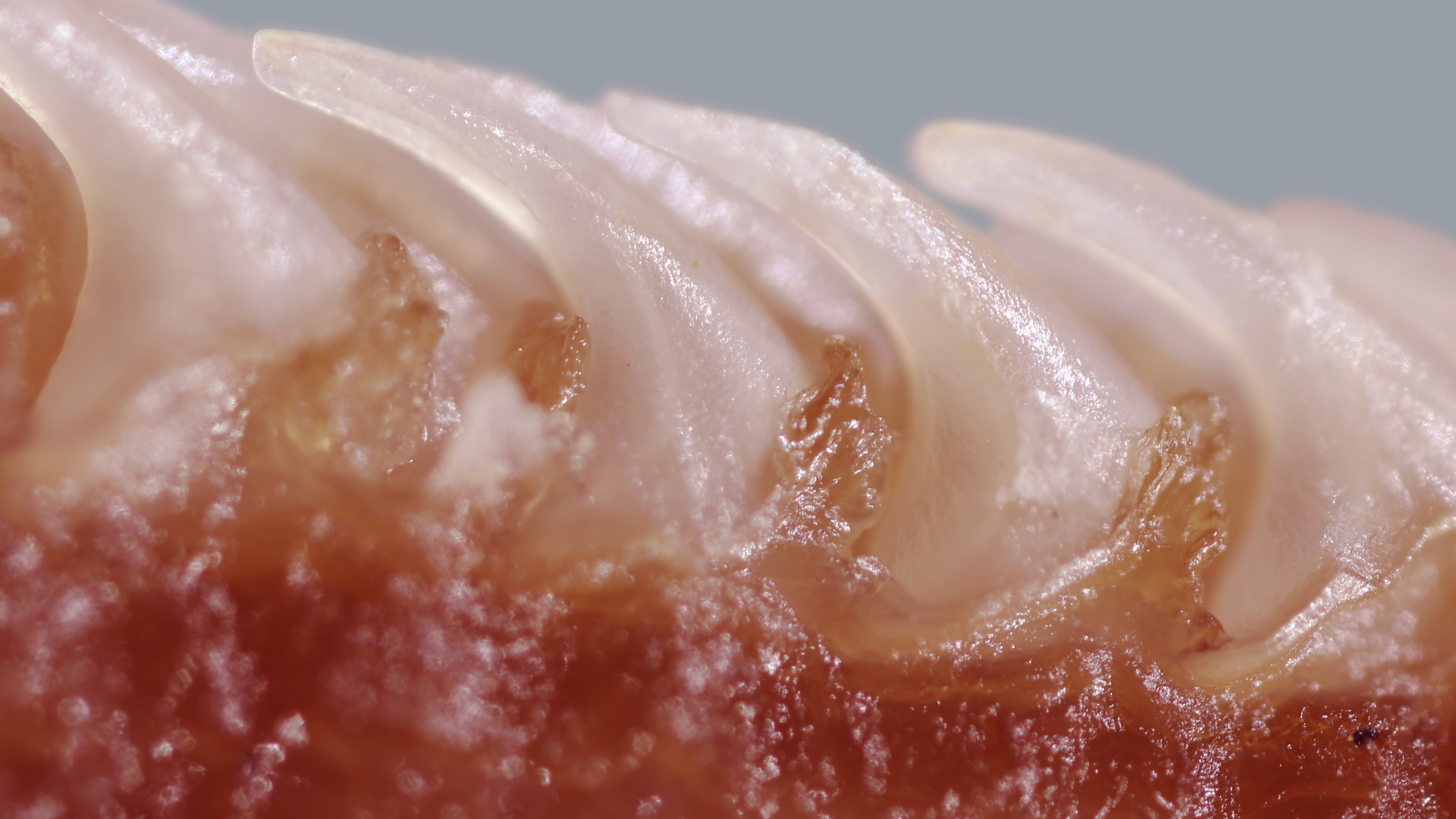
The microscopic structure of a cat's tongue helps keep its fur clean The secret is in the grooves Nov 24th 2018 T .S. ELIOT'S MYSTERY cat, Macavity, besides being a criminal mastermind able. The cat tongue is most recognized for its hundreds of sharp, backward-facing keratin spines called filiform papillae, shown in Fig. 1 A and B. A 1982 study concluded that a cat papilla has the.
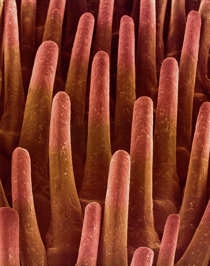
Scanning electron microscopic studies of the surface of the dorsal tongue of the cat Authors: Shin-ichi Iwasaki Hokuriku University Ken Miyata Kan Kobayashi Abstract To find out what role these spines played, mechanical engineers Alexis Noel and David Hu at the Georgia Institute of Technology in Atlanta performed microscopic computed tomography scans of the tongues of six species of cats -- the domestic cat, bobcat, cougar, snow leopard, tiger and lion.

Falsecolour SEM of the surface of a cat's tongue Stock Image P475/0027 Science Photo Library
The M P of the FuP, a sense organ for taste, of the cat tongue was considered to be a transformed type from the MP of the FiP, which acquires the ability to masticate and swallow functionally, Key words: Cat -Filiform papillae -Fungiform papillae -Microvascular structure Scanning electron microscope Introduction Many previous studies have been. A correlated light microscopic, transmission and scanning electron microscopic study of tissue samples of representative areas of the cat tongue showed that filiform papillae on the tip of the tongue were short and exhibited several conical processes from the base of each papilla. A pronounced species variation in the organization of filiform papillae has been observed.
