The 3D Cell Viewer is a tool for viewing thousands of 3D images of cellular structures and organelles. About Allen Cell Collection Data & Digital Tools Analysis & Modeling Publications Education Support 🔍 3D Cell Viewer. 3D Cell Viewer. The Institute. For example, cell polarization could be more accurate depicted in 3D cell cultures models unlike in 2D models in which the cells can only be partially polarized. Moreover, greater stability and longer lifespans were found in 3D culture models; 3D spheroids can be cultured up to 3 wk, whereas 2D monolayer culture can last for less than a week due to the limitation of cell confluence[ 33 ].

human cell 3d model
A 3D model of a Eukaryote including the major components, while missing a few smaller structures: Ribosomes and Lyosomes and a number of tiny filament. Please note, that usually the cells are so densely packed with structures, that if this was an accurate representation of the amount of components, it would be didactically unusable. Therefore it was decided to only have a few copies of each. 3D cell culture models are in vitro multicellular structures designed to emulate tissue or organ like properties that better replicate the cellular environment in vivo, providing opportunities to produce more relevant results.Both organoid and spheroid models are being increasingly used in multiple areas such as neurobiology stem cell research, regenerative medicine, and cancer biology for the. 3D cell models are increasingly being used to understand disease mechanisms and discover drug therapeutics. The process may involve deriving 3D cultures, such as cancer organoids, from patients. The 3D cultures can be used to screen for small molecule drugs or genetically manipulated to understand disease pathways. Advantages of 3D Cell Models. Three-dimensional (3D) cell models are more physiologically relevant than two-dimensional cell cultures, and they more closely represent the tissue microenvironments, cell-to-cell interactions, and biological processes that occur in vivo.Now you can generate more predictive data using high-content imaging (HCI) systems like the ImageXpress system.
10 Trendy 3D Animal Cell Model Project Ideas 2023
A general scheme of areas of 3D cell culture systems in development. The 3D cell culture systems are being developed to mimic physiological organs in the body to evaluate drug absorption, distribution, metabolism and excretion, cardiac function in the heart, and immune response in the lymphatic system. The optimal goal is to integrate these. The developing human cerebellum has a greater diversity of progenitor types than that of the mouse, necessitating a human-based model for studying cerebellar development and disease. Atamian et al.1 developed a 3D organoid model of cerebellar development, which recapitulates many cell types found in the developing human cerebellum, including Purkinje-neuron-like cells. Study shows 3D organization of DNA controls cell identity programs. by Bridget Kuehn, Cornell University. Transcriptional changes and enhancer remodeling accompany early developmental decisions. a. Animal cell made in blender for biology class. - Animal cell - Downloadable - Download Free 3D model by Lauri Purhonen (@LauriPurhonen)

Animal Cell 3D Model medical CGTrader
Orbit navigation Move camera: 1-finger drag or Left Mouse Button Pan: 2-finger drag or Right Mouse Button or SHIFT+ Left Mouse Button Zoom on object: Double-tap or Double-click on object Zoom out: Double-tap or Double-click on background Zoom: Pinch in/out or Mousewheel or CTRL + Left Mouse Button Integrate 3D models with your curriculum. Content units: Prokaryotic and Eukaryotic Cells, Monocot and Dicot Plant Structure, Energy, Genetics, Blood Cells, Animal Form and Function, Evolution and Animal Diversity; Use lesson plans to connect 3D models with learning goals and NGSS and state standards; Study the details in dozens of 3D models.
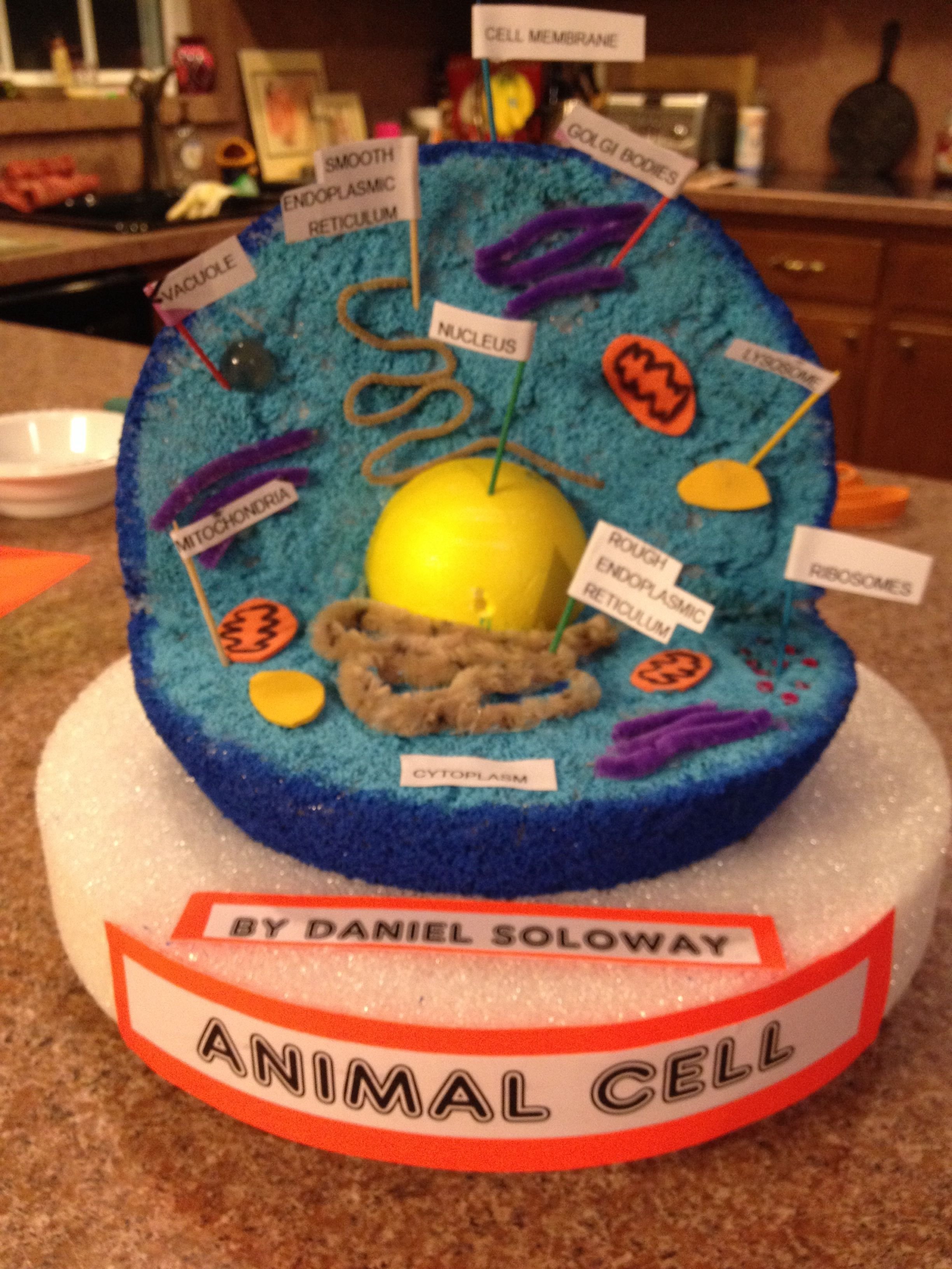
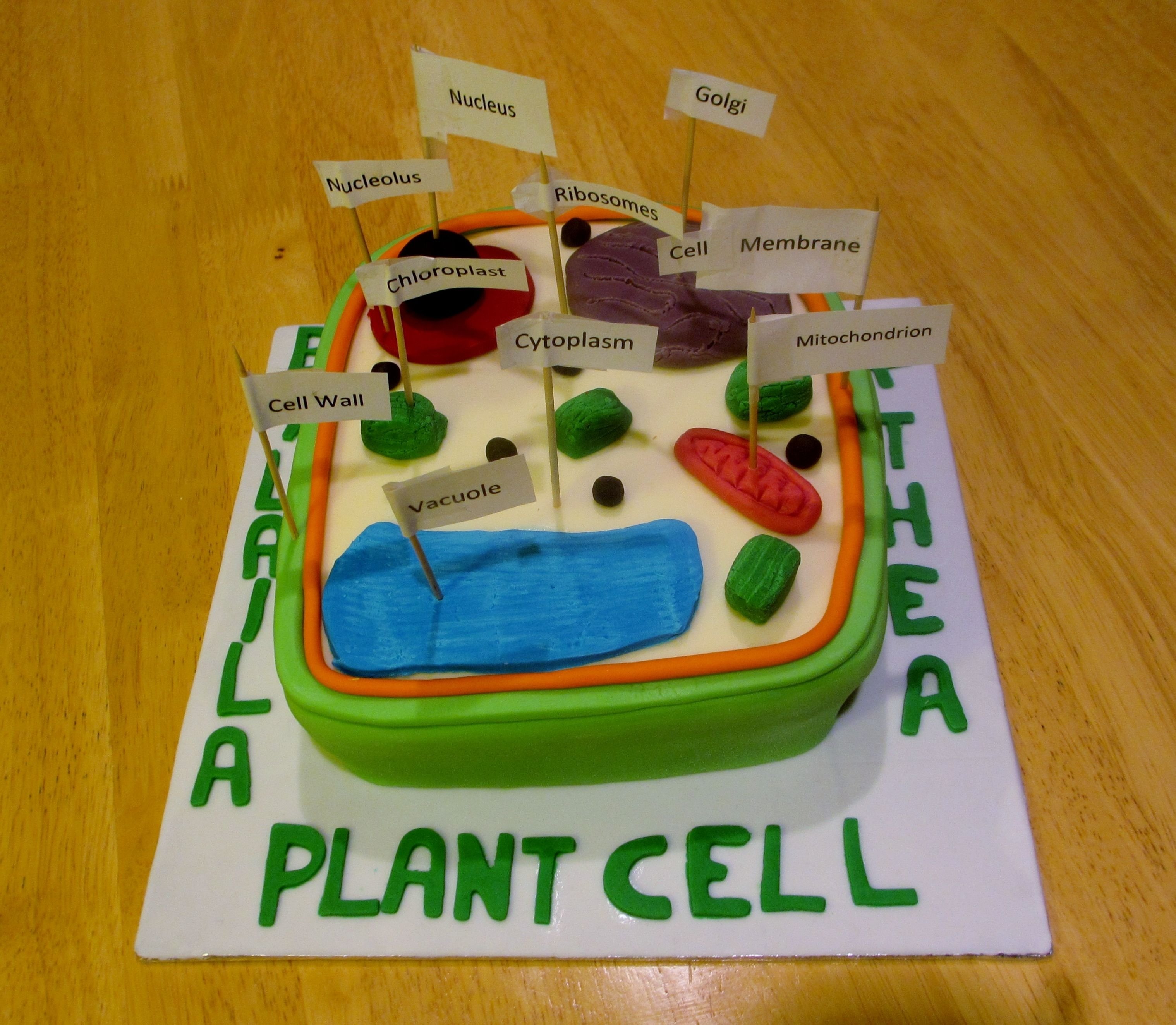
Vesicles A cytoplasmic organelle bounded by a single membrane and containing oxidative enzymes. Lysosomes are digestive animal cell vesicles that recycle worn-out cell parts, debris and food. Centrioles They are a pair of barrel-shaped organelles close to the nuclear envelope and help determine the skeletal framework of the cell by aiding in. Creating 3D models of animal and plant cells is an engaging way to deepen one's understanding of cellular structure and function. Whether you opt for clay modeling, everyday craft materials, or dive into the world of 3D printing and modeling software - each method offers a unique perspective on these fascinating microcosms that are.
10 Stylish 3D Plant Cell Model Ideas 2023
Step 3: Consider the Parts of the Cell. Now you need to make a list of all the parts, or organelles, that need to be included in your 3D cell model. Organelles are the "mini organs" that are found inside every plant and animal cell. Each organelle has a different function and physical appearance, and together they work to keep the cell alive. 5. Add the cell parts. Add the parts to your cell base (the styrofoam). This can be done by using hot glue, regular glue, toothpicks, pins, staples, or a number of other methods. In some cases you may also need to literally dig or carve out space in the styrofoam to fit in the parts.
