Anatomy The back comprises the spine and spinal nerves, as well as several different muscle groups. The sections below will cover these elements in more detail. Spine The spine is composed of. Dec. 24, 2023, 4:25 AM ET (Yahoo News) Human skeletons, remains of sharks, blood-sucking bats. human skeleton, the internal skeleton that serves as a framework for the body. This framework consists of many individual bones and cartilages.

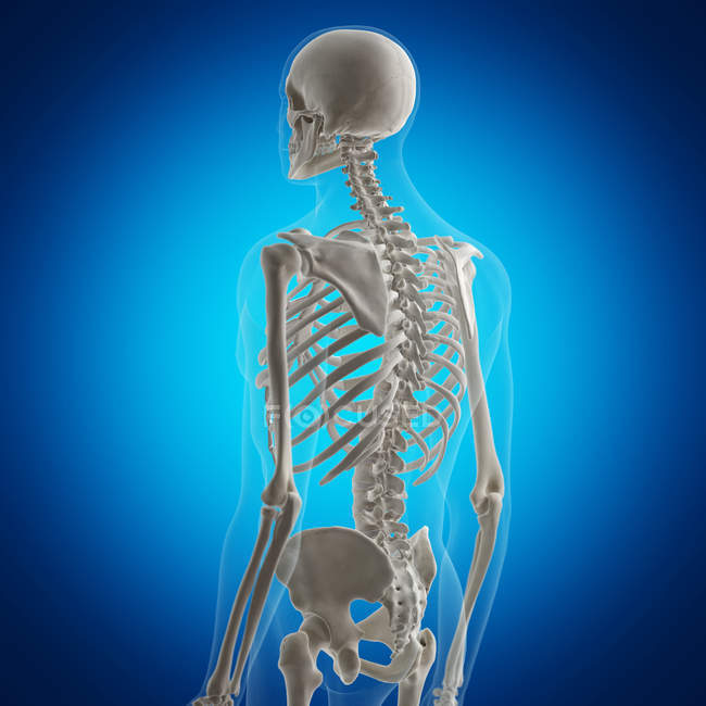
Illustration of back bones in human skeleton on blue background
The back is the body region between the neck and the gluteal regions. It comprises the vertebral column (spine) and two compartments of back muscles; extrinsic and intrinsic. The back functions are many, such as to house and protect the spinal cord, hold the body and head upright, and adjust the movements of the upper and lower limbs. Spine Anatomy Overview Video Typical Anatomical Problems that Cause Back Pain Spinal pain can arise from problems in the bones, nerves, or other soft tissues. Many of the intricate structures in the spine can lead to pain, and pain can be concentrated in the neck or back area, radiate to the extremities, or be referred to other parts of the body. The bones of the back, together, make up the vertebral column.The vertebral column is made up of 5 sections: the cervical vertebrae, the thoracic vertebrae, the lumbar vertebrae, the sacrum and the coccyx.These sections total 33 vertebrae which function together to aid locomotion and posture as well as providing support and protection. Whilst each section of the vertebral column consists of. The human back, also called the dorsum ( pl.: dorsa ), is the large posterior area of the human body, rising from the top of the buttocks to the back of the neck. [1] It is the surface of the body opposite from the chest and the abdomen. The vertebral column runs the length of the back and creates a central area of recession.

Human Bone Anatomy Back / The spine Anatomy of the spine Anatomy
The muscles of your lower back and flexibility of your lumbar spine allow your trunk to move in all directions — front to back (flexion and extension), side to side (side bending) and full circle (rotation), as well as twist. The last two lumbar vertebrae allow for most of this movement. Protects your spinal cord and cauda equina. The muscles of the lower back help stabilize, rotate, flex, and extend the spinal column, which is a bony tower of 24 vertebrae that gives the body structure and houses the spinal cord. The spine, or backbone, is a bony structure that supports your body. It connects different parts of your musculoskeletal system, which includes your body's bones and muscles. Your spine helps you sit, stand, walk, twist and bend. Advertisement Cleveland Clinic is a non-profit academic medical center. Greater Trochanter Iliofemoral Ligament Iliolumbar Ligament Ischiofemoral Ligament Joint Capsule of Hip L1 (1st Lumbar Vertebra) L2 (2nd Lumbar Vertebra) L3 (3rd Lumbar Vertebra) L4 (4th Lumbar Vertebra) L5 (5th Lumbar Vertebra) Lesser Trochanter Obturator Membrane Pelvis Posterior Sacroiliac Ligament Pubic Symphysis Pubofemoral Ligament

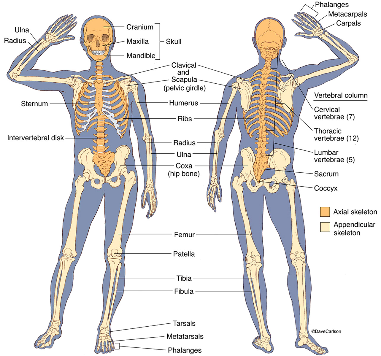
Human Skeleton Carlson Stock Art
Anatomy of the Back. A collection of articles covering the anatomy of the back, including the muscles of the back and the vertebral column. All. Latest. The skeletal system is made up of more than 200 bones and has two main parts: the axial and appendicular skeleton. Find labeled diagrams here.. Lumbar vertebrae: Five bones in the low back region; Thorax . The thorax contains the sternum (breastbone) and the thoracic (rib) cage. The thoracic cage comprises 12 pairs of ribs connecting to the.
The back is a key topographical region of the body, with crucial importance for posture, locomotion, and upper and lower limb movements. [1] The spine, located in the midline, divides the body into unequal anterior and posterior segments. In the posterior segment, the body area between the neck and gluteal regions is defined as the back region. Bones, discs, and joints in your lower back. Your lower back contains 5 vertebral bones stacked above each other with intervertebral discs in between. These bones are connected at the back with specialized joints. The lumbar spine connects to the thoracic spine above and the hips below. Individual anatomical structures include 2 Cramer GD.

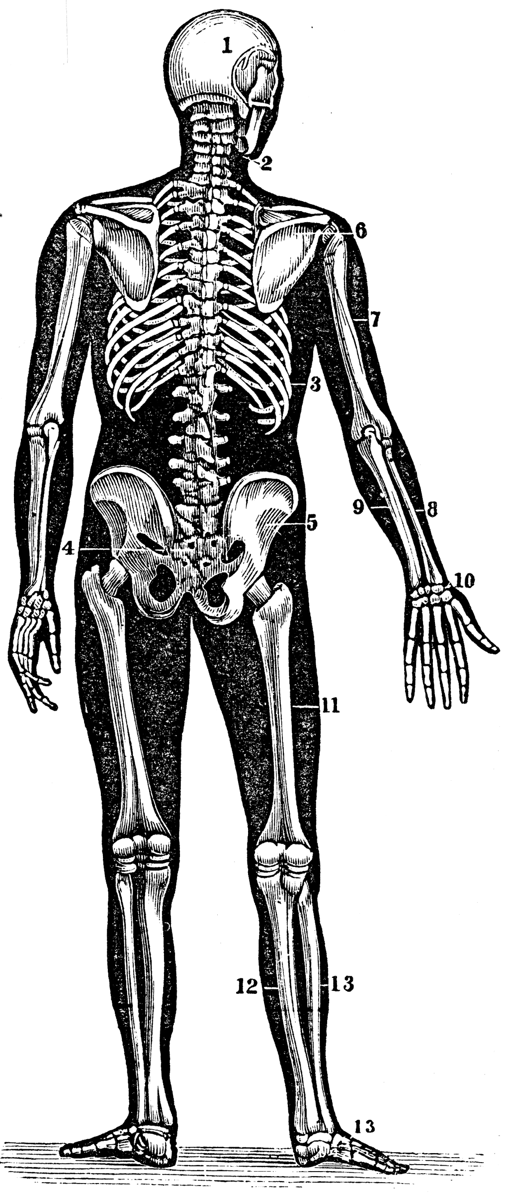
human back anatomy
Latissimus dorsi (lats), the largest muscle in the upper part of your body. It starts below your shoulder blades and extends to your spine in the lower part of your back. Levator scapulae, a smaller muscle that starts at the side of your neck and extends to the scapula (shoulder blade). Rhomboids, two muscles that connect the scapula to the spine. The Skeletal System Explore the skeletal system with our interactive 3D anatomy models. Learn about the bones, joints, and skeletal anatomy of the human body. By: Tim Taylor Last Updated: Jul 29, 2020 2D Interactive NEW 3D Rotate and Zoom Anatomy Explorer HEAD AND NECK CHEST AND UPPER BACK PELVIS AND LOWER BACK ARM AND HAND LEG AND FOOT


