Quick idea: in this article, you will learn the location of different organs from the different systems (like skeletal, digestive, respiratory, urinary, cardiovascular, endocrine, nervous, and special sense) of a dog with their important anatomical features. Dog anatomy details the various structures of canines (e.g. muscle, organ and skeletal anatomy). The detailing of these structures changes based on dog breed due to the huge variation of size in dog breeds. Would you be surprised to know that short dogs are more aggressive? Or taller dogs are more affectionate?

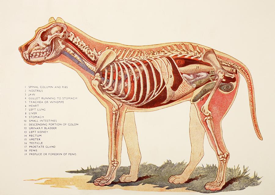
Internal Organs Of A Male Dog. From Photograph by Ken Welsh Fine Art
On the left side view of a dog's internal organs, you can see the lungs, heart, liver, stomach, spleen, kidney, intestines, bladder, and the rectum in that order from front to back. You can also view the spinal column and the brain. Laurie O'Keefe Dog Anatomy Organs Right Side Dog - Muscles Dog - Thorax/Abdomen/Pelvis Animal - Anatomy atlas: Cardiovascular system Veterinary anatomy - Animal: ANATOMICAL PARTS Abdomen Abdominal aorta Abdominal mammary gland Abdominal mammary region Accessory carpal bone Acromion Adductor muscle Ala of ilium; Wing of ilium Ala of nose Anconeus muscle Antebrachial region Aortic arch Dog anatomy comprises the anatomical studies of the visible parts of the body of a domestic dog.Details of structures vary tremendously from breed to breed, more than in any other animal species, wild or domesticated, as dogs are highly variable in height and weight. The smallest known adult dog was a Yorkshire Terrier that stood only 6.3 cm (2.5 in) at the shoulder, 9.5 cm (3.7 in) in length. The anatomy of a dog includes its skeletal structure, reproductive system, the internal organs, and its external appearance. The following paragraphs explain all these aspects in brief, along with diagrams, which will help you understand them better. External Anatomy Dogs, like all mammals, have eyes, a nose, a forehead, and ears.

Dog Internal Anatomy Poster Dog anatomy, Anatomy, Vet medicine
Common anatomical terminology Here are some common veterinary terms and their meanings: Pet senses Pets communicate in a very different way than people do. They have the same basic senses like sight, hearing, smell, touch, and taste, but they use them differently to communicate with the world. Anatomic Planes. The main planes of motion for dogs are as follows (see Figure 5-1): • The sagittal plane divides the dog into right and left portions. If this plane were in the midline of the body, this is the median plane or median sagittal plane. • The dorsal plane divides the dog into ventral and dorsal portions. The Anatomage Dog is the first highly detailed dog anatomy atlas that comprehensively features internal organs, including vascular systems and muscular-skeletal structures. Originating from real dog data, the Anatomage Dog exhibits the highest level of anatomical accuracy. All of its volumetric 3D and individual structures are segmented, users. Xiphoid region (Cranial abdominal region) Zygomatic bone. Zygomatic gland. Zygomatic region. Radiographic anatomy: labeled images in the transverse plane of a healthy dog's whole body, using tomodensitometry. Introduction to the anatomy of the skull, thorax, abdomen, pelvic cavity, muscles and blood vessels: main anatomical structures identified.

Anatomy Of Back Organs / Anatomy Male Organs in Loop Stock Footage
A dog's physical anatomy is designed to help them navigate their environment and perform various tasks. Their bodies are made up of many different parts, including their skeleton, muscles and internal organs. One of the most important parts of a dog's anatomy is their skeleton. A dog's skeleton is made up of many different bones, which provide. Have comments? Anatomy Study Resources This guide contains a variety of resources to help with the study of veterinary anatomy. Included are web resources, books (both print and electronic), and specialty resources such as flashcards. For suggestions or comments, please submit them via the Have comments tab. Anatomy No-Cost Web Resources
This module of vet-Anatomy is a basic atlas of normal imaging anatomy of the dog on radiographs. 51 sampled x-ray images of healthy dogs performed by Susanne AEB Borofka (PhD - dipl. ECVDI, Utrecht, Netherland) were categorized topographically into seven chapters (head, vertebral column, thoracic limb, pelvic limb, larynx/pharynx, thorax and abdomen/pelvis). Thighs: The upper thigh is above the knee of the hind leg. The lower thigh is beneath the knee. Stifle: The stifle, or knee, sits on the front of the hind leg. It falls in alignment with the abdomen. Hock: The hock is also known as the harsus. This is the joint on the dog's hind legs that makes an awkward sharp angle.

Глубокие мышцы, внутренние органы собаки Dog Muscles & Internal
Trachea Lungs and Smaller Airways (bronchi and bronchioles) Dog Mouth Lungs Cat Chest Dog Chest Special Senses The organs of special senses allow the animal to interact with its environment; sight, taste, smell and hearing. Cat Eyes Dog Eyes Dog Ears Urogenital System This module of vet-Anatomy presents an atlas of the anatomy of the head of the dog on a CT. Images are available in 3 different planes (transverse, sagittal and dorsal), with two kind of contrast (bone and soft tissues).


