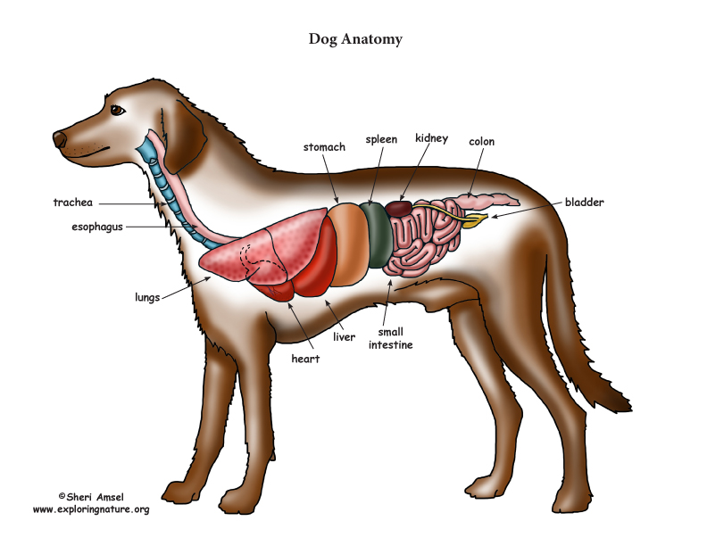
Summary Anatomy of a Dog Dog anatomy details the various structures of canines (e.g. muscle, organ and skeletal anatomy). The detailing of these structures changes based on dog breed due to the huge variation of size in dog breeds. Would you be surprised to know that short dogs are more aggressive? Or taller dogs are more affectionate? Quick idea: in this article, you will learn the location of different organs from the different systems (like skeletal, digestive, respiratory, urinary, cardiovascular, endocrine, nervous, and special sense) of a dog with their important anatomical features.

Dog Anatomy (Thoracic and Abdominal Organs)
Abdomen Abdominal aorta Abdominal mammary gland Abdominal mammary region Accessory carpal bone Acromion Adductor muscle Ala of ilium; Wing of ilium Ala of nose Anconeus muscle Antebrachial region Aortic arch Apex of nose; Tip of nose Arm On the left side view of a dog's internal organs, you can see the lungs, heart, liver, stomach, spleen, kidney, intestines, bladder, and the rectum in that order from front to back. You can also view the spinal column and the brain. Laurie O'Keefe Dog Anatomy Organs Right Side The anatomy of a dog includes its skeletal structure, reproductive system, the internal organs, and its external appearance. The following paragraphs explain all these aspects in brief, along with diagrams, which will help you understand them better. External Anatomy Dogs, like all mammals, have eyes, a nose, a forehead, and ears. Dog anatomy comprises the anatomical studies of the visible parts of the body of a domestic dog.Details of structures vary tremendously from breed to breed, more than in any other animal species, wild or domesticated, as dogs are highly variable in height and weight. The smallest known adult dog was a Yorkshire Terrier that stood only 6.3 cm (2.5 in) at the shoulder, 9.5 cm (3.7 in) in length.

Глубокие мышцы, внутренние органы собаки Dog Muscles & Internal
They have the same basic senses like sight, hearing, smell, touch, and taste, but they use them differently to communicate with the world. In general, pets have a much better sense of smell, hearing, and sight than humans. This allows them to identify odours better, to hear noises at greater distances, and to see in the dark. Perhaps the most significant is head shape. There are three main different types of head formation in dogs: Dolichocephaly: dolichocephalic dogs are those with a head which is longer than it is wide. The skull and snout are elongated and their eyes are located in a lateral position, making it difficult to see well bifocally. Internal anatomy of a dog: carnivorous domestic mammal raised to perform various tasks for humans. Encephalon: seat of the intelluctual capacities of a gog. Spinal column: important part of the nervous system. Stomach: part of the digestive tract between the esophagus and the intestine. Spleen: hematopoiesis organ that produces lymphocytes. Diaphragm: The diaphragm is the primary muscle involved in breathing. When a dog barks, it contracts the diaphragm forcefully to expel air out of its lungs and through its vocal cords. Laryngeal muscles: The laryngeal muscles control the opening and closing of the dog's vocal cords, which are located in the larynx (voice box) in the neck.

Anatomy of a male dog crosssection, showing the skeleton and internal
Thighs: The upper thigh is above the knee of the hind leg. The lower thigh is beneath the knee. Stifle: The stifle, or knee, sits on the front of the hind leg. It falls in alignment with the abdomen. Hock: The hock is also known as the harsus. This is the joint on the dog's hind legs that makes an awkward sharp angle. The Anatomage Dog is the first highly detailed dog anatomy atlas that comprehensively features internal organs, including vascular systems and muscular-skeletal structures. Originating from real dog data, the Anatomage Dog exhibits the highest level of anatomical accuracy. All of its volumetric 3D and individual structures are segmented, users.
Dogs have a third trochanter, which is the attachment site of the superficial gluteal muscle.. (From Evans HE: Miller's anatomy of the dog, ed 4, Philadelphia,. The ribs limit overall thoracic spine motion and protect internal organs. Joint Motion. The body segments of the forelimb and hindlimb are illustrated in Figures 5-3 and 5-4,. ISSN 2534-5087. This vet-Anatomy module presents an anatomy atlas of the abdomen and pelvis of the dog in CT. CT images are from a healthy 6-year-old castrated male dog. This module displays cross-sectional labeled anatomy images of the canine abdominal cavity and the pelvis on a Computed Tomography (CT) and 3D images of the abdomen of the dog.

dog anatomy Dog Care Training Grooming
Dogs also have organs that are vital to their survival, such as the heart, lungs, and liver. The anatomy of a dog can vary depending on their breed. For example, a Greyhound has a lean and muscular body that allows them to run at incredible speeds, while a Bulldog has a stocky and powerful build that makes them great for activities like weight. A sturdy pelvic limb Leg bones like the tibia and fibula Metatarsal bones Metacarpal bones Carpal bones Central tarsal bone Thoracic limb Your dog's teeth are important bones that help your dog chew, eat, and hunt. Keeping your dog's teeth healthy is important to their overall health. SUPPORT YOUR DOG'S BONE HEALTH


