A beautiful drawing of a Neuron. And it will teach you to draw the Neuron very easily. Watch the video and please be kind enough to thumbs up my videos. And. Classes of neurons Based on their roles, the neurons found in the human nervous system can be divided into three classes: sensory neurons, motor neurons, and interneurons. Sensory neurons Sensory neurons get information about what's going on inside and outside of the body and bring that information into the CNS so it can be processed.
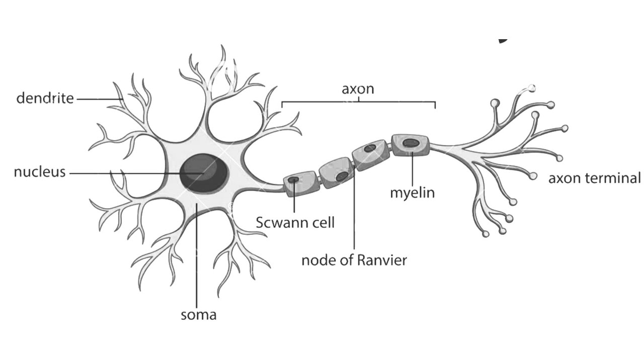
Diagram Of A Neuron exatin.info
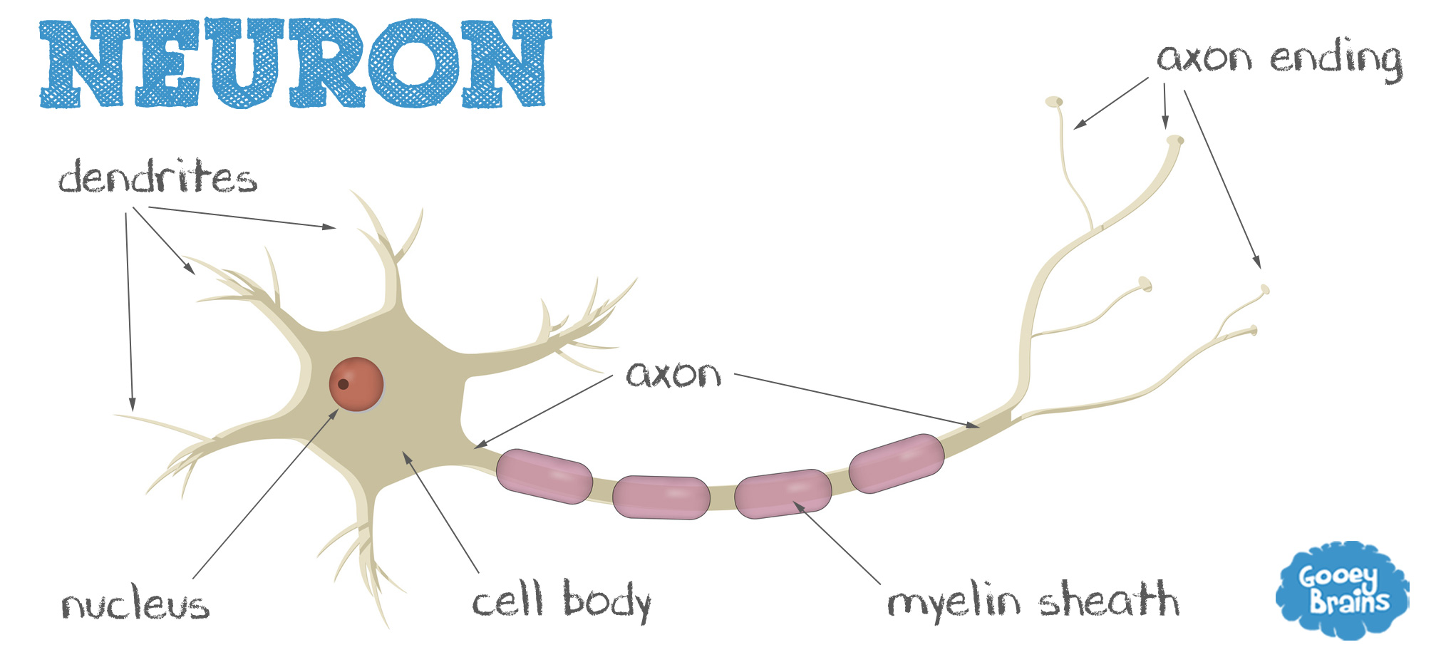
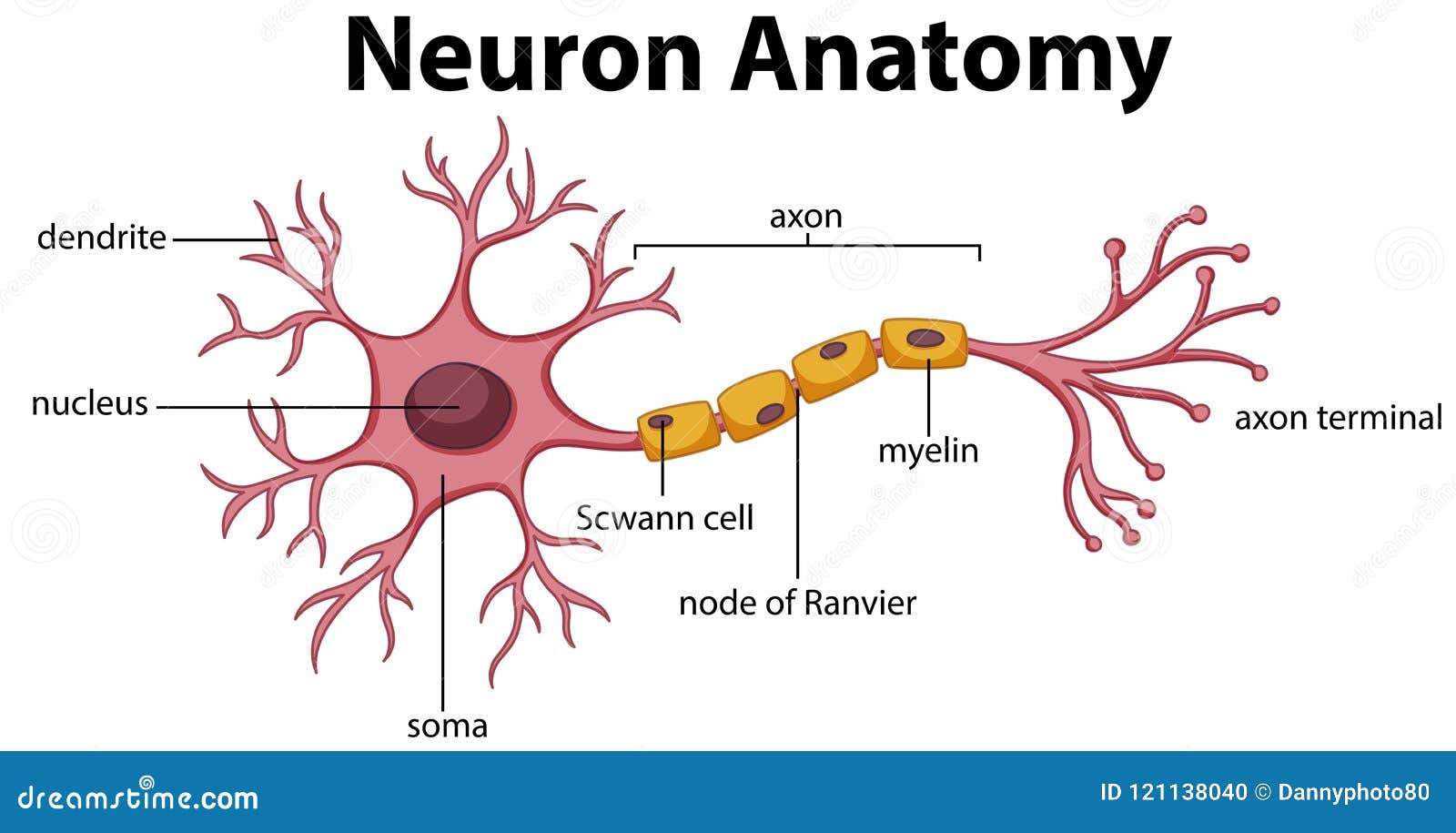
How to Draw a Neuron Manocha Academy 1.14M subscribers Subscribe Subscribed 14K views 11 months ago How to Draw a Neuron Easy! Our Website: http://bit.ly/2KBC0l1 Android App:. Quick and simple step by step drawing lesson showing how to draw a neuron (motor neuron). For more science drawing lessons visit www.ellenjmchenry.com, click on VIDEOS. Also, search. Neurons (or nerve cells) are specialized cells that transmit and receive electrical signals in the body. Neurons are composed of three main parts: dendrites, a cell body, and an axon. Neuron Anatomy Nerve Cell: Dendrites receive messages from other neurons. The message then moves through the axon to the other end of the neuron, then to the tips of the axon and then into the space between neurons. From there the message can move to the next neuron. Neurons pass messages to each other using a special type of electrical signal.

Neurons 3d illustration. Being Patient
Neuron Diagram: Direct students to complete question #3 on the Viewing the Neuron Worksheets (draw a diagram of a neuron; see Figure 2 for example drawings). Figure 2. Example student drawing of a neuron. Neuron doctrine Drawing of neurons in the pigeon cerebellum, by Spanish neuroscientist Santiago Ramón y Cajal in 1899. (A) denotes Purkinje cells and (B) denotes granule cells, both of which are multipolar. The neuron doctrine is the now fundamental idea that neurons are the basic structural and functional units of the nervous system. Axon. The axon, also called a nerve fiber, is a tail-like structure of the neuron that joins the cell body at a junction called the axon hillock. The function of the axon is to carry signals away from the cell body to the terminal buttons to transmit electrical signals to other neurons. Acting as a conduit, the axon carries these signals to. Illustration by Sophia Smith Parts of a neuron Neurons vary in size, shape, and structure depending on their role and location. However, nearly all neurons have three essential parts: a cell.
What is a neuron? Understand their vital role in the nervous system
33 By Roberta Smith Jan. 18, 2018 It's not often that you look at an exhibition with the help of the very apparatus that is its subject. But so it is with "The Beautiful Brain: The Drawings of. Every exquisite drawing by Santiago Ramón y Cajal, the founder of modern neuroscience, is marred by a curious mark. Here is the little-known story behind it. Generations of neuroscientists have wondered why the elegant and historically important drawings of Santiago Ramón y Cajal, the field's founder, are marred by a prominent blue stamp.
How TO Draw neuron cell easy/draw nervous system easyit is very easy drawing detailed method to help you.i draw the nervous system with pencil on art paper o. Neurons are electrically excitable cells that transmit signals throughout the body. Neurons employ both electrical and chemical components in the transmission of information. Neurons are connected to other neurons at synapses and connected to effector organs or cells at neuroeffector junctions. A typical multipolar neuron is comprised of soma or cell body, an axon, and dendrites.
Neuron Diagram Labeled Human Anatomy
At a synapse, one neuron sends a message to a target neuron—another cell. Most synapses are chemical; these synapses communicate using chemical messengers. Other synapses are electrical; in these synapses, ions flow directly between cells. At a chemical synapse, an action potential triggers the presynaptic neuron to release neurotransmitters. These are Spanish neuroanatomist Santiago Ramón y Cajal's drawings of neurons. Over five decades of work, Cajal (1852-1935) created more than 2,900 drawings detailing the nervous system's.
