
Human Cardiac Anatomy koibana.info Heart anatomy, Heart diagram
—The Children's Heart Institute HASAN ABDALLAH, FAAP, FAAC www.childrenheartinstitute.org SUPERIOR VENACÀVÀ PULMONARY ARTERY ra LUNG PULMONARY ARTERY The Heart this drawing shows how Olcod 'lows through the heart. Color Me. The ate.2S the heart With oxygen ate labeled with at'l Color these areas The areas o' the heart with less The heart has three layers. They are the: Epicardium: This thin membrane is the outer-most layer of the heart. Myocardium: This thick layer is the muscle that contracts to pump and propel blood. Diagram of Heart. The human heart is the most crucial organ of the human body. It pumps blood from the heart to different parts of the body and back to the heart. The most common heart attack symptoms or warning signs are chest pain, breathlessness, nausea, sweating etc. The diagram of heart is beneficial for Class 10 and 12 and is frequently. Function and anatomy of the heart made easy using labeled diagrams of cardiac structures and blood flow through the atria, ventricles, valves, aorta, pulmonary arteries veins, superior inferior vena cava, and chambers. Includes an exercise, review worksheet, quiz, and model drawing of an anterior view (frontal section) of the heart in order to.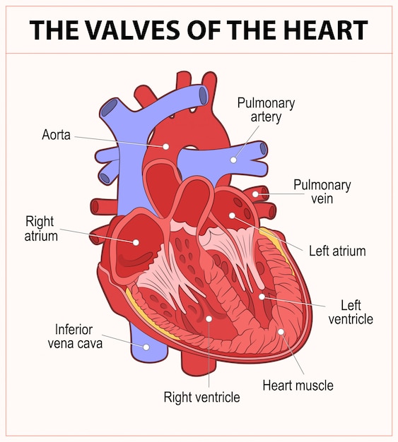
Premium Vector Diagram of human heart anatomy
Did you know that an adult human heart beats over 100,000 times a day? How many times would it beat in a year? 1 2 3 4 5 6 7 8 9 10 11 12 Color and label parts: Drag and drop the text labels onto the boxes next to the diagram. Selecting or hovering over a box will highlight each area in the diagram. Download Exercise. Tweet.. In this interactive, you can label parts of the human heart. Drag and drop the text labels onto the boxes next to the heart diagram. If you want to redo an answer, click on the. Heart Worksheet. Heart Worksheet to test your knowledge of the five basic parts of the human heart. Once you've finished labeling the diagram, color it in! You can read all about your heart and find a science project to do here. ←. heart, organ that serves as a pump to circulate the blood. It may be a straight tube, as in spiders and annelid worms, or a somewhat more elaborate structure with one or more receiving chambers (atria) and a main pumping chamber (ventricle), as in mollusks. In fishes the heart is a folded tube, with three or four enlarged areas that correspond.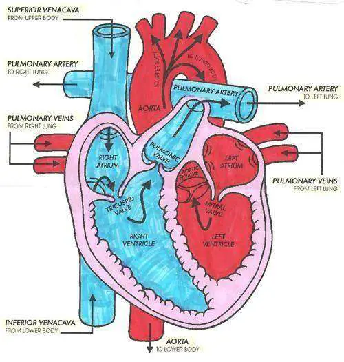
Heart diagram Healthiack
Use Crayola® crayons, colored pencils, or markers to color the human heart. Color the areas numbered 1 red. They are the arteries. Color the areas numbered 2 blue. They are the veins. Did you know?The human heart is made up of two different kinds of blood vessels. Blood vessels are hollow tubes that carry blood all over the human body. The body has three kinds of vessels: arteries. Description. This printable includes four beautifully clear and simple cross sectional diagrams of the human heart. They photocopy well and are great for use as a labeling and coloring exercise for your students. The first heart diagram is a colored and labeled reference chart. In case you need a little refresher before going over your lesson. 17,477 human heart diagram stock photos, 3D objects, vectors, and illustrations are available royalty-free. See human heart diagram stock video clips. Hand drawn illustration of human heart anatomy. Educational diagram showing blood flow with main parts labeled. Vector illustration easy to edit. Step 1 and 6 involve a blood vessel, which makes sense as this is how blood enters and exits that side of the heart. Steps 2-5 involve a chamber, valve, chamber, and valve. So if you remember this general pattern, it will help you recall the order in which blood flows through each side of the heart.

