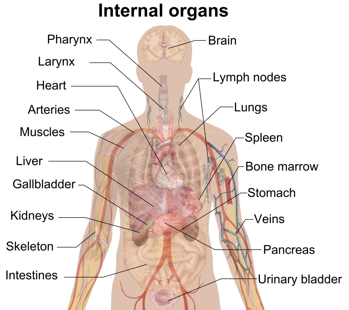
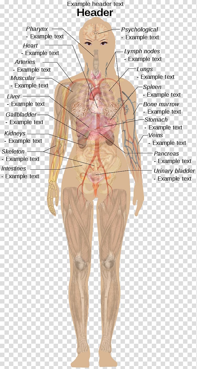
The five vital organs in the human body are the brain, heart, lungs, kidneys, and liver. Other organs include the gallbladder, pancreas, and stomach. Organ systems, such as the nervous. human body, the physical substance of the human organism, composed of living cells and extracellular materials and organized into tissues, organs, and systems. Human anatomy and physiology are treated in many different articles.

Organ (biology) Wikipedia
The Wikimedia Human body diagrams is a collection of images whose main purpose is to provide a way of explaining medical conditions and other phenomena. Contents 1 Diagrams 2 Human body diagrams 2.1 How to derive an image 2.1.1 Derive directly from raster image with organs 2.1.2 Derive "from scratch" 2.1.3 Derive by vector template Diagram External Internal Breast Anatomy Functions Female anatomy includes the internal and external structures of the reproductive and urinary systems. Reproductive anatomy plays a role in sexual pleasure, getting pregnant, and breastfeeding. The urinary system helps rid the body of toxins through urination (peeing). Key points Humans—and other complex multicellular organisms—have systems of organs that work together, carrying out processes that keep us alive. The body has levels of organization that build on each other. Cells make up tissues, tissues make up organs, and organs make up organ systems. There are 12 major anatomy systems: Skeletal, Muscular, Cardiovascular, Digestive, Endocrine, Nervous, Respiratory, Immune/Lymphatic, Urinary, Female Reproductive, Male Reproductive, Integumentary. Select a system below to get started. ANATOMY SYSTEMS Skeletal System The skeletal system includes all of the bones and joints in the body.

Image result for human organs diagram Human Anatomy Female, Human
You are here: BBC Science > Human Body & Mind > The Body Human Anatomy - Organs Click on the labels below to find out more about your organs. More human anatomy diagrams: nervous system,. An anatomy atlas should make your studies simpler, not more complicated. That's why our free color HD atlas comes with thousands of stunning, clearly highlighted and labeled illustrations and diagrams of human anatomy. No missing information, no confusion, and no hidden costs; simply a learning resource you can trust to make your studies easier. Dimensions: 674 x 599 Photo description: This diagram of the human body shows a range of organs that are important to human anatomy. They include the brain, heart, lungs, spleen, muscles, stomach, kidneys and more. This topic page will provide you with a quick introduction to the systems of the human body, so that every organ you learn later on will add a superstructure to the basic concept you adopt here. Contents Skeletal system Muscular system Cardiovascular system Respiratory system Nervous system Central nervous system Peripheral nervous system

Pin by Microlife India on Microlifeindia Medical Entrance Human body
The human body is the entire structure of a human being. It is composed of many different types of cells that together create tissues and subsequently organs and then organ systems. They ensure homeostasis and the viability of the human body. The main bones in the abdominal region are the ribs. The rib cage protects vital internal organs. There are 12 pairs of ribs and they attach to the spine. There are seven upper ribs, known as.
Use the model select icon above the anatomy slider on the left to load different models. Premium Tools. My Scenes allows you to load and save scenes you have created. All annotations, pins and visible items will be saved. Zygote Scenes is a collection of scenes created by Zygote Media Group with annotations identifying anatomical landmarks. Pancreas. Bladder. Spleen. The organs of the body chart is laminated and wipeable marker pens can be used to make notations. Our range of anatomical wall charts make perfect displays for use by professionals in a clinic and for students. Designed and printed in the UK. Organs chart dimensions: 65 cm x 50 cm.

Diagram Internal Female Anatomy Internal Organs of the Human Body
Body cavity labeled diagram of organs they contain, membranes, and lateral views. Body cavity definitions and subdivisions in tables and charts. Ventral, dorsal, cranial, spinal, vertebral, thoracic, pleural, pericardial, mediastinum, abdominopelvic, abdominal, and pelvic cavities explained. Quiz yourself with the labeled views! Human Body Diagrams INDEX Musculoskeletal Skeleton & Spine Shoulder & Back Arm & Hand Pelvis & Hip Leg & Foot Circulatory Nervous Digestive Urinary Reproductive Medical Art Library is a resource for teachers, students, health professionals or anyone interested in learning about the anatomy of the human body. We are medical artists who love anatomy.


