Over 90% Of All Products On eBay Are Brand New. Big Brands, Top Retailers. Great Prices On Millions Of Items. Get It On eBay. Read the latest neuroscience research. Submit your work to eLife's new model of publishing. Following review, revise, resubmit or declare Version of Record - it's up to the author

Unlabeled Brain Diagram Wiring Diagram
Labeled brain diagram. First up, have a look at the labeled brain structures on the image below. Try to memorize the name and location of each structure, then proceed to test yourself with the blank brain diagram provided below.. Unlabeled (blank) diagram to be used as a worksheet Learn faster with quizzes. Learn all about brain. Next, here are 3 unlabeled human brain diagrams. Students can print these and practice labeling the parts of the human brain in preparation for a psychology, biology, or anatomy quiz or test. Blank Human Brain Diagram. Finally, here is a set of blank human brain diagrams. These may be useful for teachers to include in posters. The Brain; The Brain - Map Quiz Game. Cerebellum; Cerebrum; Frontal lobe; Occipital lobe; Parietal lobe; Spinal cord; Temporal lobe; You need an account to play. Create challenge. 0/0 0 % Game mode: Pin Type Show more game modes. Learn. Restart---Your high score (Pin) Log in to save your results. A topographical anatomy of the brain showing the different levels (encephalon, diencephalon, mesencephalon, metencephalon, pons and cerebellum, rhombencephalon and prosencephalon) as well as a diagram of the various cerebral lobes (frontal lobe, occipital, parietal, temporal, limbic and insular). Please note that the limbic lobe is functional.
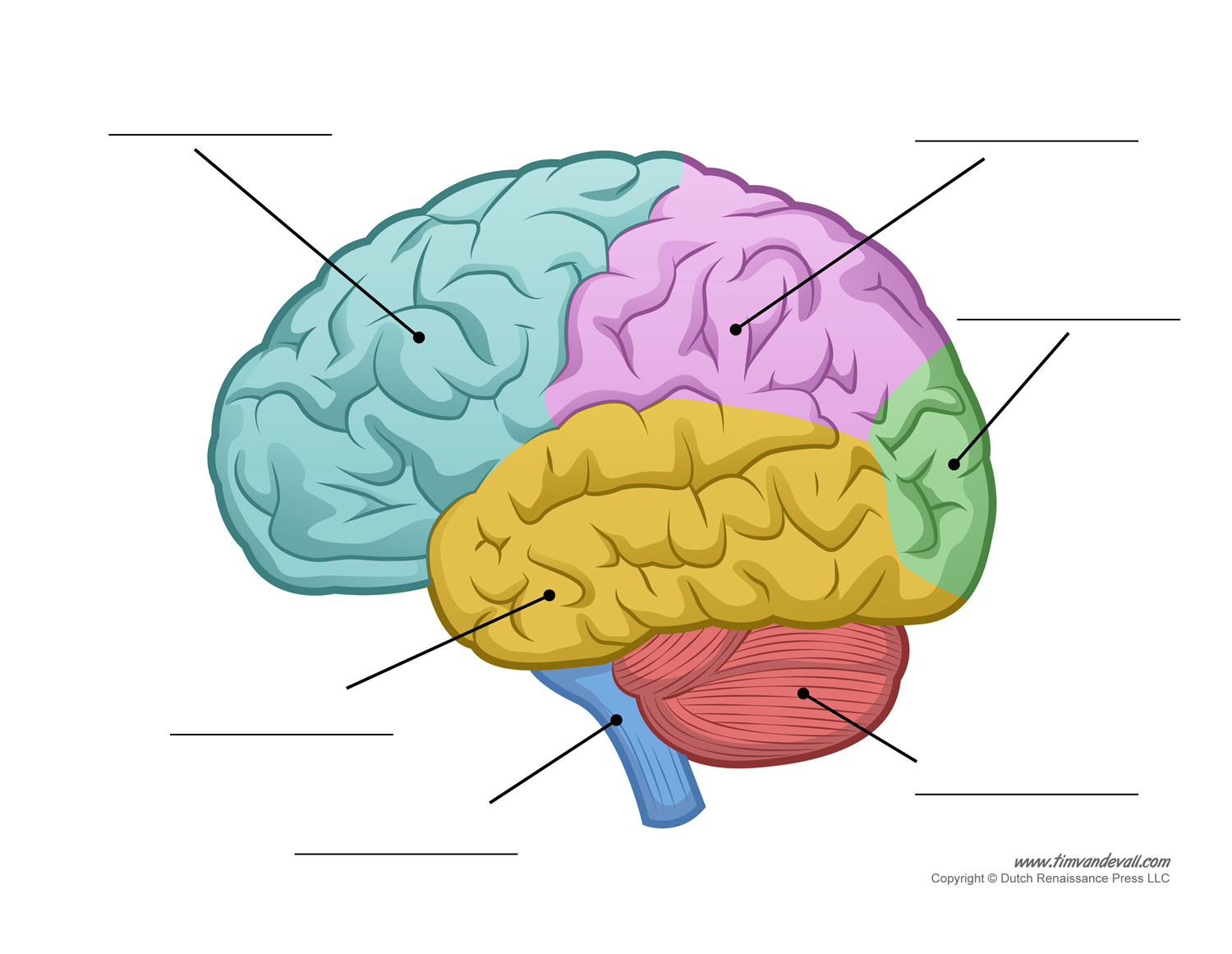
Brain Diagram Unlabeled Color Tim van de Vall
Brain diagram without text.svg. From Wikimedia Commons, the free media repository. File. File history. File usage on Commons. File usage on other wikis. Metadata. Size of this PNG preview of this SVG file: 800 × 571 pixels. Other resolutions: 320 × 228 pixels | 640 × 457 pixels | 1,024 × 731 pixels | 1,280 × 914 pixels | 2,560 × 1,828 pixels. The midsagittal section of the brain shows the three major parts of the brain, which are the cerebrum, cerebellum, and brainstem.The cerebrum (prosencephalon or forebrain) comprises the telencephalon (cerebral hemispheres) and the diencephalon.They are each also divided into subparts or regions for simplified localization of structures, for example, the brainstem is composed of the midbrain. What's In Your Brain? Activity Key 1. Cerebral cortex 2. Thalamus 3. Corpus callosum 4. Hypothalamus 5. Hippocampus 6. Pituitary gland 7. Midbrain 8. Pons 9. Medulla 10. Brainstem 11. Spinal cord 12. Cerebellum Clicking here to how a free human brain image. Teaching the parts by this human brain with these convenient printables by students and teachers. Skip in content. Principal Menu. Home; Library (2023-2018) Library (2015-2017) Publish; Over Menu Toggle. Contact; Join; Login Menu Toggle. Account; Purchase History; Edit Your Profile; Cancelation;
Unlabeled Brain Diagram Cliparts.co
Our collection includes a variety of brain diagrams, ranging from simple and labeled diagrams for beginners, to more detailed and unlabeled diagrams for advanced learners. Each diagram highlights key structures and areas of the brain, such as the frontal lobe, parietal lobe, occipital lobe, temporal lobe, and cerebellum, among others. Image size: 31.2 Mpixels (89.4 MB uncompressed) - 6250x5000 pixels (20.8x16.6 in / 52.9x42.3 cm at 300 ppi) Published in: Brain and Nervous System Images , Neurology Images & Videos. Powered by PhotoDeck.
In Part 2 of Brain Matters: Brain Diagram, students fill in the names of structures and functions on an unlabeled brain map. They check accuracy by comparing their work with other students. Parts 1 and 2 can be completed independently of each other. Goals and Objectives Students will be able to: • identify the functions of structures of the brain Unlabeled Nervous System Images. The images shown below are not labeled in any way and are taken using different powers of microscope magnification. You should study the previous chapters covering specific tissues of the nervous system to give you the knowledge you need to determine the type of tissue (s) or structures shown in each picture.

The Wired Mind—VIRGINIA Magazine
We introduce the Mindboggle-101 dataset, the largest and most complete set of free, publicly accessible, manually labeled human brain images. To manually label the macroscopic anatomy in magnetic resonance images of 101 healthy participants, we created a new cortical labeling protocol that relies on robust anatomical landmarks and minimal manual edits after initialization with automated labels. Given below is a diagram of brain, its functions, detailing the four lobes and their associated structures. Four Lobes of the Brain. Frontal Lobe. This is the part of the cerebrum that lies directly below the frontal bone. It is the part that is present directly behind the forehead. It is separated from the parietal lobe by the central sulcus.
