EFFECTIVE LEARNING CENTRE THE BEST HUMAN BODY PICTURES
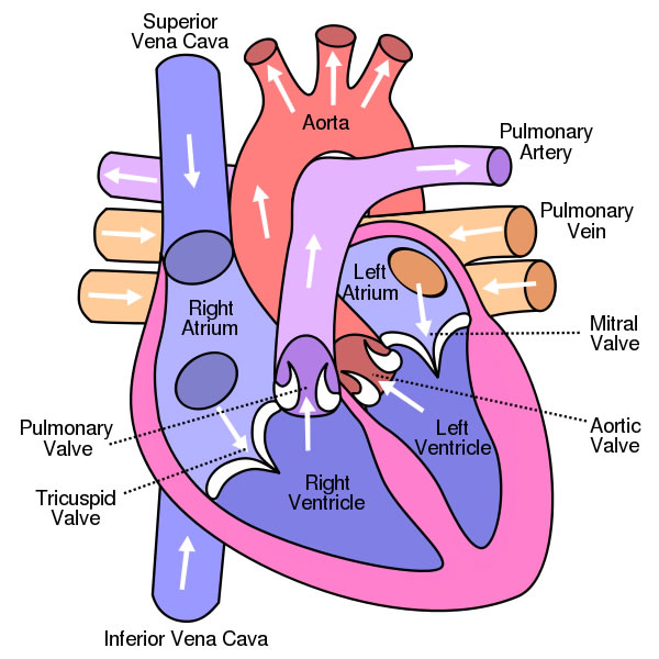
Welcome to the anatomy of the heart made easy! We will use labeled diagrams and pictures to learn the main cardiac structures and related vascular system. In addition to reviewing the human heart anatomy, we will also discuss the function and order in which blood flows through the heart. Don't forget to LABEL the parts of the heart on the diagram! 1. Compare the location of the tricuspid and bicuspid. 2. Compare the direction of blood flow in the pulmonary artery to the pulmonary vein. 3. Mitral regurgitation is a heart condition that occurs when the mitral valve does not close fully. Heart. Your heart is the main organ of your cardiovascular system, a network of blood vessels that pumps blood throughout your body. It also works with other body systems to control your heart rate and blood pressure. Your family history, personal health history and lifestyle all affect how well your heart works. The heart is located in the thoracic cavity medial to the lungs and posterior to the sternum. On its superior end, the base of the heart is attached to the aorta,mycontentbreak pulmonary arteries and veins, and the vena cava. The inferior tip of the heart, known as the apex, rests just superior to the diaphragm.
Human Cardiac Anatomy koibana.info Heart anatomy, Heart diagram
heart, organ that serves as a pump to circulate the blood.It may be a straight tube, as in spiders and annelid worms, or a somewhat more elaborate structure with one or more receiving chambers (atria) and a main pumping chamber (ventricle), as in mollusks. In fishes the heart is a folded tube, with three or four enlarged areas that correspond to the chambers in the mammalian heart. Did you know that an adult human heart beats over 100,000 times a day? How many times would it beat in a year? 1 2 3 4 5 6 7 8 9 10 11 12 Color and label parts: This printable includes four beautifully clear and simple cross sectional diagrams of the human heart. They photocopy well and are great for use as a labeling and coloring exercise for your students. The first heart diagram is a colored and labeled reference chart. Diagram Of Heart Diagram of Heart The human heart is the most crucial organ of the human body. It pumps blood from the heart to different parts of the body and back to the heart. The most common heart attack symptoms or warning signs are chest pain, breathlessness, nausea, sweating etc.
Color heart diagram CardiacExercise Nursing school notes, Medical
Use Crayola® crayons, colored pencils, or markers to color the human heart. Color the areas numbered 1 red. They are the arteries. Color the areas numbered 2 blue. They are the veins. Did you know?The human heart is made up of two different kinds of blood vessels. Blood vessels are hollow tubes that carry blood all over the human body. The body has three kinds of vessels: arteries. Step 1 and 6 involve a blood vessel, which makes sense as this is how blood enters and exits that side of the heart. Steps 2-5 involve a chamber, valve, chamber, and valve. So if you remember this general pattern, it will help you recall the order in which blood flows through each side of the heart. Sometimes it helps to see how something works rather than read about it. That's why the Heart Institute at UPMC Children's Hospital of Pittsburgh added this to the Family Guide to Your Child's Heart Care. Ask your child's heart doctor to use these diagrams to explain how a normal, healthy heart works. Then ask the doctor to draw how your child. Download and print these Anatomy Heart coloring pages for free. Anatomy Heart coloring pages are a fun way for kids of all ages to develop creativity, focus, motor skills and color recognition. Anatomy Coloring Pages - Heart. Find the Resources You Need! Search . More Teaching Resources:

